Reasons to choose List Labs as your Microbiome CDMO:
We support a very diverse offering in Bacterial Manufacturing.
We are a passionate and transparent CDMO.
We are flexible and cohesive enough to be an extension of our customer’s team.
We offer end-to-end solutions for our customers.
Learn more about List Labs’ 45 years of experience with Bacterial Manufacturing by watching our video below:
This interview between Jessica Thompson from Kisaco Research and Stacy Burns-Guydish, PhD, President of List Labs, talks about the growth of the Microbiome field for Microbiome CDMOs and digs deep into the Live Biotherapeutics side of this burgeoning industry.
To get further insight into the future of the Microbiome field, check out the video below:
Jessica Thompson (Kisaco Research)
Hi, and welcome to the latest of our interview spotlights in the run-up to Microbiome Connect USA. My name is Jessica Thompson. I’m the Portfolio Director of the Life Sciences and Microbiome Portfolio for Kisaco Research. And it’s a pleasure to be joined by Stacy Burns- Guydish, who is the president at List Labs. Stacy, thank you so much for joining.
Stacy Burns-Guydish, PhD, President (List Labs)
Well, thanks for having me.
Jessica Thompson (Kisaco Research)
So to start off Perhaps, it would be great if you could give a little bit of an introduction about yourself and your background in the space.
Stacy Burns-Guydish, PhD, President (List Labs)
Yeah, so I’m the president of List Labs and my background is in microbiology. I got my PhD at Baylor College of Medicine and then did my postdoc at Stanford. A lot of my early microbiome career was focused on more on infectious disease of bacteria and that host-pathogen interaction. Before I joined List Labs, I joined a startup company that was using a strict anaerobic clostridium organism to make a biofuel or a biochemical. So, we improved this organism for making that biochemical, and we scaled the fermentation process up to 100,000 liters. So, this was a really good experience that I brought to List labs and working with strict anaerobes and that scale-up of that fermentation and immediately applied it to Live Biotherapeutic Products (LBPs) and that process development. So, since I’ve been at List Labs now for over six years, I’ve been involved with many of the LBP companies and moving their products to clinical trials.
Jessica Thompson (Kisaco Research)
Great. and what excites you about working in microbiome research
Stacy Burns-Guydish, PhD, President (List Labs)
So, I’m going back a little bit in my history. When I was in high school, in my first biology class the first time, I looked under a microscope, and I was hooked. I knew that I wanted to be a microbiologist, and I knew that I wanted to do something that would benefit society. But I think what I didn’t realize then is how beneficial those organisms in the microbiome could be to our health. You know, bacteria tended to get a bad rap. They’ve always been thought of as ones that cause infection, but they are important to our health. The research in the Microbiome field is revealing all these important synergies that these bacteria serve in our bodies. So, they helped develop our immune and endocrine systems. They help us digest our food, they provide vitamins and nutrients, and they also prevent and protect us from infection. So, it’s exciting to watch the growth in this field from more uncharacterized donor derived products. And now we’re seeing more characterized single organisms or a consortium of bacteria that are being used as a therapeutic to improve patient lives. The field is also evolving from there. So, we’re seeing engineered organisms that can deliver a therapeutic to a specific site. Also, we’re seeing you know a kill switch inserted into the organism, so they provide their function and then they’re eliminated. Then we’re also seeing companies go on to identify the metabolites and the proteins that are also derived from this microbiome associated bacteria.
Jessica Thompson (Kisaco Research)
Yeah, I think it’s an exciting field so far. There’s been so many developments in the field over the past few years for this. Given all the excitement, there’s still obviously a lot of challenges to overcome within the space. What do you think are some of the major challenges that are facing the LBP industry or the microbiome industry more broadly?
Stacy Burns-Guydish, PhD, President (List Labs)
I think there’s two major challenges for me, one is that you know this is a unique product, this LBP Live bacterial organism. So that organism’s viability needs to be maintained through the whole entire manufacturing process. So, this can be a hurdle for some organisms that are difficult to grow. You know these strict anaerobes that oxygen is toxic to them, right? It impacts their viability, and this becomes even more of an issue as you scale up these organisms. It’s one of the reasons why you know people want to probably partner with a Contract Development Manufacturing Organization (CDMO). CDMOs understand what those hurdles are, they know about what you’re going to encounter as you scale up these organisms. So, they can help you optimize that process and knows how to overcome those hurdles as part of the scale up.
I think the second the biggest challenge in the industry is the investment arena. They’re being cautious. There’s no commercial product approved yet, so everyone’s waiting for that BLA approval from you know either fairing or series. And then once that happens, I think that’s really going to drive the field forward.
Jessica Thompson (Kisaco Research)
Yeah, I would agree for sure. And turn into List Labs a little bit more. So, if you’re going to hear a bit about what your company sort of focus in the space your expertise is and how you’re hoping to help overcome those challenges really.
Stacy Burns-Guydish, PhD, President (List Labs)
List Labs was started in 1978. So, we have over 40 years of experience with bacteria. We started with bacterial-derived products. And we have over 100 products in our catalog of high-quality reagent-grade products used in medical research and vaccine development. We also have the experience that we designed and built our facility specifically for containment and segregation to be able to do GMP manufacturing. Also, it was designed for the use of spore formers. So, we have a long history of working with all different kinds of organisms. Strict anaerobes, aerobic organisms, and spore formers. We also have the experience performing GMP manufacturing for many different companies that have gone on to late-stage clinical phase and who now have commercial products.
So, you know, we have quite a bit of history of moving companies through their clinical phase to success to commercial. So, with all this experience that we have, it fits well with the microbiome space. Because we have experience with all those different organisms, we are one of the first companies to produce and manufacture a live biotherapeutic that went into clinical trials. Since then, we’ve manufactured dozens of LBPs that have gone into clinical trials. We’ve also just recently installed a 500-liter fermenter here at our facility. So, we can continue to support companies through their phase 2 manufacturing when they need larger capacity.
Jessica Thompson (Kisaco Research)
And, of course, you partnered with genome and company last year to further your capabilities even further than that. Can you speak to what motivated this partnership and how you’re hoping it will be beneficial over the coming years?
Stacy Burns-Guydish, PhD, President (List Labs)
Yeah. So, List Labs here at the California facility has served customers for Phase 1 and Phase 2 manufacturing, but we aspired to provide our customers with an end-to-end solution. We also saw that there was an opportunity that there wasn’t a lot of capacity in the market for commercial LBP Manufacturing. Genome And Company also had a large pipeline of LBPs that they also wanted to secure their early and late phase manufacturing for. So, this was really a very synergistic relationship with List Labs and Genome And Company. List Labs had the expertise we’d manufactured LBPs and have GMP experience. We have experience designing our own facility, while Genome And Company have very interested investors that are ready to build a new facility for commercial manufacturing.
Jessica Thompson (Kisaco Research)
It sounds like it will be a really exciting development to watch. And with that. And just for this lab, I suppose as well, what are your ambitions then for the next 5, 10 years? What are you hoping for in terms of your next steps?
Stacy Burns-Guydish, PhD, President (List Labs)
You know, so it’s all about supporting our customers that ended in a solution. So, we’re planning to build a new facility in fishers, Indiana, for Phase 3 and commercial manufacturing. That facility is going to be 130,000 square feet specifically for LBPs. It will have four manufacturing lines, and it will be able to accommodate all different types of organisms, from aerobes to strict anaerobes and spore formers.
The production lines will include a 2000-liter fermenter and it will either be stainless steel or single use type fermenter to provide flexibility for our customers. We’re also going to have the ability to formulate and fill in a strict anaerobic environment which we really believe is a differentiator for us. In this facility, we also have QC laboratories, office space, and GMP warehouse space as well. And our ambitions are to expand from there. You know, we want to have sites around the world and we see ourselves as a global Microbiome CDMO company.
Jessica Thompson (Kisaco Research)
I’ll be watching this space then to see where you’re emerging over the next few years. So, we’re really excited to be welcoming you as a platinum sponsor at Microbiome Connect next month in Boston. What are you looking forward to about attending the conference?
Stacy Burns-Guydish, PhD, President (List Labs)
List Labs is super excited about being a part of this conference and to support and advance the microbiome field. I’m really looking forward to the amazing agenda that Kisaco Research has put together. I’m looking forward to hearing updates regarding emerging therapeutics and specifically about Microbiome derived metabolites that might be used in autism as a therapy.
We’re also excited to hear about gut microbiome candidates that improve the efficacy of checkpoint inhibitors for cancer. Also, how companies are advancing their process strategies, such as co-culturing for consortium organisms and single-use alternatives as well. We’re going to hear about engineered organisms for specific delivery of a therapeutic to a site and also great talks from regulatory and quality representatives as well.
So, I think this conference is going to be excellent, not just for a seasoned microbiome veteran but also for those new scientists and innovators that are coming into the field.
Jessica Thompson (Kisaco Research)
Yeah, it was a real pleasure putting together the program this year. There’s so much exciting work that’s being done in the space. It’s almost too much to choose from in terms of when it comes to putting together the talks, and obviously, you’re going to be presenting as part of our bio-processing strategies track as well as chairing that track of the conference in terms of your presentation. Can you give us a quick snippet for the audience in terms of the key takeaways you’re hoping they’ll get from your presentation?
Stacy Burns-Guydish, PhD, President (List Labs)
So, I think we’re going to do something a little bit different than what other CDMOs have previously done. You know, you’re going to get to hear a success story from one of our actual clients, Sciota Biosciences. They have a groundbreaking phase one clinical trial that they’ve just completed using a unique activated bacterial therapeutic for autism. We worked alongside Sciota to develop their process and specifically to develop and manufacture a unique vehicle that’s a part of their drug product. This project was truly a partnership, and I think it’s a great example of List Lab’s ability to be a partner. We’re transparent, and we want to drive to innovate, solve problems and provide solutions.
Stacy Burns-Guydish, PhD, President (List Labs)
Sciota’s story is inspirational, and it’s really going to tug on your heartstrings a little bit. But, I think it enforces, at least, what motivates me, and I think others and their passion for advancing LBP therapies for a healthier world.
Jessica Thompson (Kisaco Research)
Great, well, I definitely look forward to listening, and I think it’s going to be a great addition to the program. So, thanks so much for taking the time to talk to me. It’s great to hear a little bit more about List Labs, and I look forward to seeing what you guys all do in the future. Thanks so much.
Stacy Burns-Guydish, PhD, President (List Labs)
Thank you so much for your time
List Labs is an experienced contract development and manufacturing organization (CDMO) harnessing bacteria’s potential for a healthier world. We offer a refreshingly flexible approach to clients seeking a CDMO for Live Biotherapeutic Products (LBP) derived from beneficial bacteria. Partner with List Labs as your CDMO and experience our uniquely collaborative and transparent culture. With over 40 years of experience, our expertise encompasses aerobes, anaerobes, and spore formers, process development and scale up, analytical development and QC testing, aseptic filling, purification, lyophilization, and cGMP manufacturing for clinical trials and future commercial launch. List Labs produced the first LBP for clinical trial before LBP was even an acronym, followed by dozens of products that led to successful clinical LBP programs. List Labs provides a solid foundation in science, purpose-built state-of-the-art facilities, and a passionate team to ensure the success of your LBP project.
New CDMO Services Trade Show Exhibit booth and Trade Show Updates
In June, we unveiled our new services trade show exhibit at the Microbiome Movement Drug Development Summit 2022. This new trade show exhibit highlights our expanded service offerings and experience in this field.
Breakdown of our new services trade show exhibit
Our company mission statement:
Harnessing Bacteria’s Potential for a Healthier World
Expanded Services:
cGMP Manufacturing Phase 1, 2, 3 & Commercial
Live Biotherapeutics
Bacterial Products
MCB & WCB
Drug Substance
Drug Products
Process Development
1L to 2000L
QC Testing
Analytical Development
Stability
Aerobes
Anaerobes
Spores
BSL1-3
Along with Fermentation, Purification, Fill, and Lyophilization services.
We then highlight our experience and the life of our company by sharing the year (1978) we started List Labs.
Next year at the Microbiome Movement Drug Development Summit 2023, we’ll upgrade our exhibitor package and extend our presentation time to introduce List’s expansion and the addition of late phase and commercial manufacturing capacity.
We recently purchased and installed our Sartorius 500L Stedim bioreactor and already have client projects booked and scheduled. Please contact us to discuss scheduling your project! Our new 500L SUB provides the capacity your project need as you progress toward late phase and commercialization.
New supporting equipment has also been purchased and installed. The AKTA filtration system will support the significant increase in scale while maintaining the high quality and purity our customers expect.
Our vision is to take our partners from pre-clinical and early phase trials to the commercialization of their products. This new equipment is helping that vision become a reality!
End-to-End LBP Manufacturing Services
Now that we have started the development of our manufacturing facility in Fishers, Indiana, we are close to genuinely becoming an end-to-end LPB Manufacturing organization, offering early-stage development, Phase I, II, III, and Commercial Manufacturing.
Contact us if you have any questions or would like to partner on your next project!
Anthrax toxin can enter living cells and the toxin enzymes, Lethal Factor (LF), and Edema Factor (EF) make known changes. Because of this activity, anthrax toxins are valuable tools to investigate cell processes. Some of the work currently accomplished with these toxins can be described by the following selected references:
Work with anthrax toxins is providing a better understanding of cellular processes. Protectative Antigen (PA) binds specifically to two toxin receptors, tumor endothelial marker 8 (TEM8, also called ANTXR1) and capillary morphogenesis 2 (CMG2, or ANTXR2). Several other factors are involved in the internalization of anthrax toxin.
Abrami L, Bischofberger M, Kunz B, Groux R, van der Goot FG (2010) Endocytosis of the Anthrax Toxin Is Mediated by Clathrin, Actin and Unconventional Adaptors. PLoS Pathog 6(3): e1000792. PMID: 20221438
Anthrax vaccines are currently under development and demonstration that antibodies will neutralize anthrax toxin is essential.
Laws TR, Kuchuloria T, Chitadze N, et al (2016) A Comparison of the Adaptive Immune Response between Recovered Anthrax Patients and Individuals Receiving Three Different Anthrax Vaccines. PLoS One11(3):e0148713. Published 2016 Mar 23. doi:10.1371/journal.pone.0148713 PMID: 27007118
PA could potentially deliver polypeptides and compounds to the cell cytoplasm. In the two studies described in the following papers, PA was used as a tool to deliver biochemicals to the cytoplasm of eukaryotic cells.
Dyer PDR, Shepherd TR, Gollings AS et al (2015) Disarmed anthrax toxin delivers antisense oligonucleotides and siRNA with high efficiency and low toxicity. J. Control. Release 2015, 220, 316–328. PMID: 26546271
Rabideau, AE, Liao XL, Akcay G, Pentelute BL (2015) Translocation of Non-Canonical Polypeptides into Cells Using Protective Antigen. Sci Rep 5: 11944, PMID:26178180, PMCID: PMC 4503955
PA has been used to target cancer cells overexpressing TEM8. This receptor has been shown to be upregulated during tumor angiogenesis and provides a convenient target for anti-angiogenic therapy.
Chen KH, Liu S, Bankston LA, Liddington RC, Leppla SH (2007) Selection of anthrax toxin protective antigen variants that discriminate between the cellular receptors TEM8 and CMG2 and achieve targeting of tumor cells. J Biol Chem 282: 9834–9845. PMID: 17251181, PMCID: PMC2530824
Chaudhary A, Hilton MB, Seaman S, et al (2012) TEM8/ANTXR1 blockade inhibits pathological angiogenesis and potentiates tumoricidal responses against multiple cancer types. Cancer Cell 21:212–226. PMID: 22340594 PMCID: PMC3289547
You don’t need to buy an entire batch of lipopolysaccharide or wait months for production and release – Saves you valuable time
Paying for as few as 50 vials of a proven product and purchasing as you need it, while supplies last, also saves you a tremendous amount of money
Proven quality, product in use worldwide in clinical trials
Get to Phase one quickly with available GMP products
Time is money. The more time spent waiting for your materials to be manufactured is time that you are not conducting your clinical trials. Contracting a manufacturer to produce GMP products can take a year or more for manufacture and release, but with a GMP compliant product in stock, you can purchase what you need when you are ready.
Help your budget with available GMP products
There are so many costs associated with research studies and clinical trials, who wouldn’t want to save a little money on their project? The expense of custom manufacturing can be steep – purchasing available GMP compliant products from List Labs can help alleviate some of those costs. Lipopolysaccharide is broadly used in many types of clinical trials such as in the study of tumor Ag-loaded IL-12 secreting semi-mature DC for the treatment of pediatric cancer.1
Lipopolysaccharide currently in use in clinical trials worldwide
List Labs’ GMP compliant version of HPT™ Lipopolysaccharide from Escherichia coli O113 has already been used by organizations around the world in clinical trials. The quality of this product is proven by the successful use in Phases one through three in past or ongoing clinical trials. This unique difference sets our GMP product apart from the competition and saves you the risk of an unknown product.
Contact us today to get your GMP LPS while there’s still product in stock!
See how List Labs’ products have been used in research projects on our citations page.
Reference
Dohnal AM, Witt V, Hügel H, Holter W, Gadner H, Felzmann T. Phase I study of tumor Ag-loaded IL-12 secreting semi-mature DC for the treatment of pediatric cancer. Cytotherapy. 2007;9(8):755-70. Epub 2007 Oct 4. PMID: 17917887
I’d like to tell you about some of the obstacles you may encounter as you develop your live biotherapeutic product (LBP) and how List Labs can help you navigate through them.
Harnessing bacteria’s potential for a healthier world is our company mission. Our history and experience have been devoted to bacteria – what bacteria can produce – cultivating bacteria, purifying proteins, and polysaccharides. List Lab’s passion is to support innovators with quality bacterial products for research and development of vaccines and medical products and to perform contract development and manufacturing service for transformative therapies such as LBPs.
List is a privately held, woman-owned, and operated company in California, and about a quarter of our staff has advanced degrees. Initially, the core part of our business was manufacturing bacterial products. Beginning with selling one bacterial product in 1978, we now have over 100 stock products, including a GMP product. List Labs is a GMP-compliant facility, but we’re more than a collection of state-of-the-art equipment, List is much greater than the sum of its parts. Live biotherapeutic projects are not a cookie-cutter process and we are uniquely qualified to provide insight and flexibility to match the needs and requirements of each individual project. We typically take on 2 to 3 microbiome projects at a time, providing your project with individual attention and the critical advantage necessary to achieve a successful outcome.
Our cGMP compliant facility has 7 expertly designed manufacturing suites allowing segregation of product campaigns and spore containment if needed. The suites undergo treatment with vaporous hydrogen peroxide prior to GMP manufacturing to ensure the quality of your product. Due to the design of the facility and equipment options, we have the ability to manufacture several products in parallel.
Leveraging decades of experience cultivating a variety of microorganisms and GMP manufacturing experience for our products and partners, the transition to the microbiome space was a natural extension of our capabilities and expertise. We began a project about 7 years ago with a partner to develop and manufacture a live bacteria product. One of the first in the burgeoning microbiome field.
We have now worked on dozens of projects for indications in the gut, skin, women’s vaginal and urinary tract health, and CNS. We manufacture products for Phase I and II clinical trials. And we have produced over 20 different LBP products. These projects not only include manufacturing but also typically require a lot of development, many of which come straight from the academic bench with very little development past a shake flask or bottle cultivation. We are a partner who is invested in the success of your project and work as an extension of your team.
Let’s use climbing Mt Everest as an analogy. We are climbing the mountain with you, right alongside you. It’s a great analogy because it gets harder and harder the higher you get, and what you do at the bottom, your preparation will make or break you at the top. Aggressive timelines, budget constraints, these are the shear cliffs we can see but so many of the obstacles that await are unseen black ice, or bottomless crevasse’s… I know it’s a bit dramatic but getting a LBP to market is much the same, full of potential pitfalls.
So, today I want to share with you what some of these pitfalls look like and give you a glimpse of how List Labs can help you avoid becoming one of the many, that never realize the ultimate goal.
Source of Strain
If there is anything you take home from this talk, it should be this. “Choose Wisely!” Your choice of strain is made early in the project and changing mid-ascent even to “quote” the same species has a compounding impact. If you need to change strains, all of your in vivo and in vitro assay will need to be repeated along with all your development. The result can be very costly and cause a substantial delay to your timeline. A strain identified as a particular species is not equal to another strain identified as the same species. Each is unique and the characteristics or phenotypes that are important for your strain and indication may not be representative of another strain although identified as the same species.
But whatever your strain of choice is, it is highly likely that we have worked with it or a similar strain before. This is a sample list, although not exhaustive, of the strains we have worked with and of course not giving away any confidentiality of our clients.
Animal-Free Media Replacement
Another potential obstacle is replacing the media with animal-free alternatives. Since you need to establish a product that is BSE/TSE-free, many clients choose to switch to animal-free media. This is not trivial as there are many animal-derived components that are difficult to replace and are necessary for the robust growth of the organism. Shown in the graph below, is an animal-based media compared to an animal-free base media missing a critical animal-derived component which when the client came to us they had been unsuccessful in replacing. Growth in an animal-free media may impact growth rate, final cell density, phenotype (which may be an important characteristic for your indication), potency, and viability. All of which has implications to the scale of the process in order to have enough viable cells for your dose requirements. This directly impacts your Return on investment.
But we know how to tackle the problem. This is an example using the graph I showed you before where growth rate and final cell density were impacted without a very critical animal-derived component. Once we identified a suitable replacement, we demonstrated similar or better growth than with the animal-derived media. Then we realized further improvements in cell density with process improvements resulting in a 5-10X improvement in cell density.
Preserving Viability
Another pitfall is loss in viability of your organism through the process. This is a typical manufacturing process flow for a live biotherapeutic product. Initially, the strain is cultivated in a seed culture either anaerobically or aerobically and inoculated into the large vessel (such as a stainless steel bioreactor or single-use bioreactor), then harvested by tangential flow filtration, formulated, lyophilized, sieved, and then filled into vials, applicators, or formulated to fill capsules.
Different organisms will have different sensitivities to the process that may impact its viability including the growth phase at which the strain was harvested, the harvest process, and the environment during the harvest, or during the lyophilization, sieving, and encapsulation process. Understanding the viability of the organism throughout the process is important to know where to focus your development efforts.
We understand these risks and what tools we can use to improve the yield of the live organism at the end of your process. We have demonstrated substantial improvements in viability by 2 to 100 fold by optimizing these variables.
Scale Up
Another hurdle to overcome is the scale-up of the process. We often start with a process that is at the tube, bottle, or shake flask scale which requires a substantial amount of scale-up development. The process development of the cultivation and harvest should be performed in a scaled-down version of the process such that performance will be predictable at scale with the necessary process controls to demonstrate similar performance. List has 1L bioreactors and scaled-down versions of the harvest process so we can isolate specific variables and understand the impact on the process. We typically perform 10X scale up from 1L to 10L to the 100L process to minimize the surprises at scale and provide a robust process. Your timeline and costs for development should include these activities.
QC Analytical Development
An obstacle of the QC Analytical Development of your LBP is the development of the bioburden assays, USP<61>/<62> which is not straightforward with a live organism. For LBPs these assays need to be tailored and developed for each organism and we have experience working on these assays. We are also able to harness a lot of efficiencies for the customer by not only working on the manufacturing but also working on the analytical development in-house. Analytical assays are necessary for both in-process testing and final QC testing for release.
Is List Labs the right CDMO for your project?
As a novice climber, would you attempt to scale Mt. Everest with anything less than the most experienced Sherpa? We are a passionate and dedicated team with the expertise and experience that is critical to reach the summit. We have been there many times. We recognize the obstacles and while no two ascents are exactly the same, we have the flexibility to guide you to the top and deliver a quality product for the success of your project. We look forward to working with you!
Check out the Overcoming Obstacles in the development of Live Biotherapeutic Products video!
Botulinum Toxin Infographic
Diphtheria Toxin Infographic
Pertussis Toxin Infographic
On May 14, 2020, a team spanning the University of California San Diego, San Francisco General Hospital, Cook County Health and Hospitals System in Chicago, and Washington University in St. Louis published a milestone study in the New England Journal of Medicine. Cohen et al demonstrated the effectiveness of LACTIN-V, a new Live Biotherapeutic Product (LBP) created by Osel, Inc. for the treatment of bacterial vaginosis (BV).
LACTIN-V is a single-strain, topically-administered LBP containing Lactobacillus crispatus CTV-05, a protective human vaginal bacterium that helps to combat the pathogenic bacteria and dysbiosis observed in recurrent BV and urinary tract infections. Its success marks the first single-strain LBP to show clinically significant efficacy in a randomized, double-blind, placebo-controlled Phase 2b trial in the US.
The study was supported by grants from the National Institute of Allergy and Infectious Diseases (NIAID), part of the National Institutes of Health (NIH). List Labs is excited by this landmark discovery, by the prospects of this discovery for women’s health around the world, and for further supporting the use of LBPs as a viable therapeutic. We had a chat with Osel’s Director of Product Development, Tom Parks, to find out more about the project, it’s challenges, and why the product is successful:
What are dysbiosis and BV?
Dysbiosis is a disruption of the human microbiome: the collection of microorganisms, including bacteria, that naturally live on and in our bodies. The full function of the microbiome isn’t fully understood, but dysbiosis is known to be involved in a wide range of skin disorders, intestinal problems and gum diseases, among many others.
Bacterial vaginosis is an ecological disorder of the vaginal microbiota in which hydrogen peroxide-producing lactobacilli are displaced by predominantly anaerobic bacteria (e.g. Prevotella and Mobiluncus species), Gardnerella vaginalis, and Atopium vaginae. It is the most common vaginal infection worldwide among women of reproductive age, and affects about 30% of women in the US. Treatment usually consists of a course of an antibiotic such as metronidazole lasting about five days. The treatment itself is usually effective, but in about 75% of cases BV returns within a year, often within just a few weeks.
Why is treating BV so challenging?
The tendency of BV to recur is due to failure to re-establish a Lactobacillus-dominated vaginal microbiome. Since the only treatment is another course of metronidazole or a similar antibiotic, this can lead to a cycle of re-infection. In addition to the discomfort and negative impact on the quality of life, BV is a risk factor for a wide range of sexually transmitted infections, including HIV/AIDS. It is also a risk factor for premature birth and other reproductive health complications.
Currently there is no approved treatment to prevent the recurrence of BV. Women have tried a range of home remedies, from yogurt to tea tree oil. As might be expected, these are often ineffective, and may have side effects of their own.
Osel has developed a treatment for BV that breaks the cycle of dysbiosis. LACTIN-V is a live biotherapeutic product (LBP) containing the hydrogen peroxide-producing strain Lactobacillus crispatus CTV-05, which is a protective member of the native microbiome. Administered after the normal course of antibiotics (once a day for five days, and then twice a week for 10 weeks), LACTIN-V helps to restore the normal vaginal environment and prevent re-emergence of the organisms that cause BV. In the study reported in NEJM, BV recurrence was significantly less in women treated with Lactin-V (30%) compared to the placebo group (45%) (P=0.01).
What goes into manufacturing an LBP?
An LBP is a therapeutic agent based around a live microorganism, intended to restore a balance in the microbiome disrupted by a disease condition. A contract development and manufacturing organization (CDMO) can provide essential expertise for developing an LBP, but finding the right CDMO is tricky since many contract manufacturing organizations do not have experience with maintaining viability of the microorganism through the entire manufacturing process or are flexible for the process nuances of an LBP product. In addition, many organisms of interest as LBPs are anaerobic to some extent (for example, Lactobacillus are facultative anaerobes), and/or spore-forming. Working with such organisms takes special expertise and facilities to provide necessary containment and segregation, maintain viability, maximize yield, and avoid pitfalls.
Osel’s Tom Parks explains, “Relevant live biologic product experience and GMP manufacturing capabilities are obviously important. Flexibility to handle a number of product formats is also helpful. Since the field is relatively new, companies that manufacture LBPs are few and far between.”
Are you looking for a CDMO for your LBP project? List Labs has 40 years of experience manufacturing bacterial products and many different LBP products for Phase 1 and 2 clinical trials for indications in the gut, skin, vaginal mucosa, and CNS. List Labs has the expertise for handling and cultivating anaerobic bacteria and spore formers. We are uniquely qualified to provide insight and the flexibility to match the needs and requirements of each individual project, such as filling different product formats. We are a passionate and dedicated partner who works as an extension of your team to ensure the success of your project. If you are interested in an LBP project, contact us at sales@listlabs.com
The purity of your recombinant proteins is critical to the success of your research. A number of contaminants can affect both in vitro and in vivo systems. Some of the results are predictable, but others are not. All can affect your experimental data.
This article will focus on one of the most common contaminants: endotoxins or lipopolysaccharides. List Labs provides certificates of analysis showing the very low levels of endotoxins in our products.
What Are Endotoxins?
Lipopolysaccharides, also known as endotoxins, are a class of complex hydrophobic molecules found in the cell membranes of Gram-negative bacteria such as Escherichia coli. They are released in large quantities following cell death and during cell division, so they are a common component of recombinant protein production.
The general structure of an endotoxin is one or more Lipid A molecules bonded to one end of a short polysaccharide oligomer. The oligomer has polysaccharide side chains that carry O-antigen. Endotoxins are generally not inactivated by heat and must be removed during purification.
Endotoxin Effects
Endotoxins have a variety of deleterious effects on mammalian systems. These can vary widely even in similar systems. In fact, at least one case is known in which two insensitive T-cell lines were cloned from an endotoxin-sensitive parent line. One factor seems to be the presence of the CD14 receptor protein on the surface of the affected cells. Higher expression of CD14 seems to correlate with greater endotoxin sensitivity. In especially sensitive systems, even picomolar concentrations of endotoxin can lead to anomalous experimental results.
Effects in vitro
Documented effects in vitro include:
Distortion of cell membranes by interactions with Lipid A, up to and including cell death.
Lesser membrane distortions still affect gene expression and protein production, with specifics varying by different types of cells.
Stimulation of leukocyte cultures to produce tissue factors.
Inducing production of IL-6 in equine macrophages.
Activating primary human immune cells, especially CD1c+ dendritic cells, to produce inflammatory cytokines. Sensitive cells can be activated by endotoxin concentrations as low as 0.02 ng/ml.
Enhancing production of prostaglandins, acid phosphatase, fibrinolytic inhibitor, collagenase, nerve growth factor, and adhesion molecule 1, depending on the cell type.
Inhibiting angiotensin-converting enzyme activity, synthesis of proteoglycan, and synthesis of alpha2 macroglobulin.
Effects in vivo
In live animals, endotoxins produce an inflammatory response in almost all tissues that are exposed to them. The pyrogenic nature of endotoxins produces effects ranging from fever to fatal septic shock.
All of the above effects, both in vivo and in vitro, may be synergistic with other contaminants or (in live animals) endogenous products. This, combined with widely varying cell sensitivity, make the experimental effects of endotoxin contamination difficult to predict.
Purification to Remove Endotoxins
The standard method of purifying recombinant proteins and removing endotoxins is affinity chromatography, using affinity tags on the target proteins, eluting the bound target, and then cleaving the tags in post-processing. Affinity chromatography is the method of choice at List Labs and gives our recombinant products exceptional purity.
Conclusion
Endotoxin contamination is a potentially serious problem in recombinant proteins, with highly variable and difficult to predict experimental effects. Even low levels of contamination may produce anomalous effects, which may vary across different cell lines and test subjects. Only reliable purification can prevent contamination. List Labs provides certified products with known purity and very low levels of endotoxin.
Dawson, “The Significance of Endotoxin to Cell Culture and Biotechnology;” LAL Update, March 1998, Vol 16, No. 1, p. 1-4; Associates of Cape Cod Incorporated; https://www.acciusa.com/pdfs/newsletter/updt0398.pdf
Schwarz, M. Schmittner, A. Duschl, and J. Horejs-Hoeck, “Residual Endotoxin Contaminations in Recombinant Proteins Are Sufficient to Activate Human CD1c+ Dendritic Cells;” PLoS One. 2014; 9(12): e113840; https://www.ncbi.nlm.nih.gov/pmc/articles/PMC4257590/
The List Labs Citation Database is a robust tool for researchers. It offers thousands of papers showing how List Labs products are used experimentally. Researchers can search keywords specific to their fields of study and learn how others in that field have used our toxins and antigens.
In this article, we will explore experimental applications for two of our most popular products: Pertussis Toxin (Products #180, #181, and #184) and Cholera Toxin (Product #100B). This article is not exhaustive – we will focus on recent research – but it offers a survey of ways in which List Labs is helping to get science done.
Experimental autoimmune encephalomyelitis (EAE) is an induced autoinflammatory condition of the central nervous system. It is used in rodents as a model of demyelinating diseases such as multiple sclerosis and of T-cell-mediated autoimmune disease in general. Inducing EAE usually uses isolated myelin proteins or homogenate along with pertussis toxin to open the blood-brain barrier and allow T-cells access to the CNS. Many citations in the database note the use of those products.
Cholera toxin was even used to study the cellular mechanisms of Yersinia pestis, the pathogen that causes bubonic plague.
This is only a brief survey of recent research using two of List Labs’ more popular products. It illustrates both the wide range of applications for List’s bacterial toxins and the utility of the List citation database as a tool to facilitate your literature surveys.
What is Coronavirus (COVID-19)?
Coronavirus (COVID-19) is a disease caused by a virus (SARS-CoV-2) that can trigger what doctors call a respiratory tract infection, and is transmitted from person-to-person.
What are the Symptoms of Coronavirus (COVID-19)?
Coronavirus (COVID-19) symptoms can range from mild to severe. Though, elderly people and people with underlying health conditions like heart or lung disease, or diabetes seem to be at a much higher risk of developing severe illness from COVID-19.
Recent Research: Experimental Applications of Pertussis Toxin and Cholera Toxin
The List Labs Citation Database is a robust tool for researchers. It offers thousands of papers showing how List Labs products are used experimentally. Researchers can search for keywords specific to their fields of study and learn how others in that field have used our toxins and antigens.
In this article, we will explore experimental applications for two of our most popular products: Pertussis Toxin (Products #180, #181 and #184) and Cholera Toxin (Product #100B). This article is not exhaustive – we will focus on recent research – but it offers a survey of ways in which List Labs is helping to get science done.
Pertussis Toxin Applications
Experimental autoimmune encephalomyelitis (EAE) is an induced autoinflammatory condition of the central nervous system. It is used in rodents as a model of demyelinating diseases such as multiple sclerosis and of T-cell-mediated autoimmune disease in general. Inducing EAE usually uses isolated myelin proteins or homogenate along with pertussis toxin to open the blood-brain barrier and allow T-cells access to the CNS. Many citations in the database note this use of those products.
Cholera toxin was even used to study the cellular mechanisms of Yersinia pestis, the pathogen that causes bubonic plague.
This is only a brief survey of recent research using two of List Labs’ more popular products. It illustrates both the wide range of applications for List’s bacterial toxins and the utility of the List citation database as a tool to facilitate your literature surveys.
List Labs has been making botulinum toxins for research for 30 years.
In that time, we’ve made reagent and GMP products for researchers and pharma companies. Our products are of the highest quality and purity; they are produced in consistent processes, tested and stabilized by freeze-drying. Proteins that need to be activated by proteolytic cleavage (nicking) are activated and purified, providing consistent proteins for your research. You do not loose toxin in the nicking process, nor do you run experiments with toxin having a variable amount of nicking. We offer both pure neurotoxin and toxin complex, fully activated.
For work with the enzymatic light chains, we offer recombinant Light Chains in four different serotypes, LC/A, /B, /C and /D which are non-toxic and may be applied to research using common laboratory practices. Recombinant heavy chain, binding domains, both GST tagged and un-tagged are available. Our toxoids are made from purified neurotoxin types A and B to give you the most specific antibody production.
We have demonstrated the use of our type A toxin antibodies; one polyclonal raised against the heavy chain is an effective capture antibody for ELISAs and other detection strategies and the other antibody, a mouse monoclonal will specifically bind to type A light chain. This antibody pair will capture and identify small amounts of toxin.
The table below lists the Product #’s for these research reagents; several are offered in different sizes.
TOXINTYPE
COMPECE
NEUROTOXIN
TOXOID
CHAINS
ANTIBODIES
A
128
130
133
611, 612, 613
730, 731
B
138
138
139
620, 622, 623
C
625
D
146
630
E
140
141
If you have questions, please contact us at sales@listlabs.com for more information.
If you are an existing customer, you can place your order with a purchase order at orders@listlabs.com
If you are not yet a customer, fill out the customer app, once approved, we can fill your order.
See information outlining the purchase of controlled toxins on our website.
Orders of Select Agent products must total less than 1mg. There are no such limitations on antibodies or chains.
We are honored to supply researchers worldwide with highly purified bacterial toxins that can potentially be instrumental in helping to change the world!
In this post, we’ve gathered all of our current citations for our Diphtheria product group. Please use these citations as a reference and resource for your potentially life-changing work!
Diphtheria Toxin & CRM
Corynebacterium diphtheriae is a Gram-positive, bacterium that infects epithelial cells of the upper respiratory tract and produces diphtheria toxin. Diphtheria toxin is proteolytically cleaved forming a two-part toxin, held together by a disulfide bridge. The amino-terminal carries the toxin’s enzymatic activity, capable of ADP-ribosylation and inactivation of translation elongation factor 2 (EF-2). The carboxy-terminal domain binds to specific host receptors, the heparin-binding EGF-like growth factor (HB-EGF) on human epithelial cells, and translocates the catalytic domain into the cell. After binding to the cell receptor, the diphtheria toxin is taken up by endocytosis, the pH of the endocytic vesicle drops, and the translocation region of the toxin helps guide the catalytic domain into the host cytoplasm where it is released. Within the cytoplasm, the diphtheria toxin catalytic domain ADP ribosylates EF-2, terminating protein synthesis and causing the death of the cell. Diphtheria toxin is highly potent, and as little as one catalytic domain is thought to cause cell death. In cell culture, diphtheria toxin inhibits protein synthesis and causes death in cells carrying the HB-EGF receptor. This toxin has been used to specifically eliminate receptor-expressing cells in transgenic mice.
Tetanus toxin (TeNT) and botulinum toxin (BoNT) appear quite different at first glance, however, when we take a closer look at how these toxins function, they are more similar than suggested by the diseases they cause.
Symptoms of Tetanus vs. Symptoms of Botulism
While tetanus causes a body to take a rigid, inflexible state, a very well-described and feared disease since antiquity; botulism reveals itself in limp, uncontrolled muscles, symptoms that mimic those of other diseases, hiding the cause of the disease until the modern age.
Symptoms are strikingly opposite: tetanus is characterized by unrelieved tension or spasticity of the striated muscles and botulism by a limp or flaccid state of the same muscles. In both cases, the muscles can no longer be moved in a coordinated manner, resulting in respiratory paralysis and death.
Tetanus and BotulismHave Similar Basic Origins and Structures
Both diseases can be attributed to toxins created by Clostridia. In general, TeNT is formed by bacteria introduced through injuries such as puncture wounds, placing the bacteria where they can grow in the absence of oxygen. BoNT is also synthesized by bacteria growing under lack of oxygen; however, in contrast to TeNT, botulinum is usually encountered when bacteria multiply and produce toxin in contaminated foods and the toxin is swallowed with the contaminated food. Both toxins enter the bloodstream and are distributed through the body (1, 2, 3, 4).
Botulinum and tetanus neurotoxins are both large proteins composed of two parts, a heavy chain, and a light chain. The light chain represents the active component; it is a protease that cleaves peptides regulating exocytosis of neurotransmitters, rendering the nerve unable to communicate. The heavy chain navigates the toxin into target cells and is responsible for transfers through several membranes.
Although botulinum and tetanus toxins have the same basic structure, tetanus neurotoxin exists solely as a two-part protein neurotoxin; where botulinum toxin is, at least initially, associated with accessory proteins, forming a toxin complex. This complex can be more than four times larger than the neurotoxin alone (5, 6).
TeNT is taken up by peripheral cholinergic nerve endings and is transported intraaxonally, retrogradely into the soma of the nerve cell (7). It leaves the motor neuron and subsequently enters nerve endings of inhibitory interneurons (8, 9, 10, 11). Within the inhibitory neurons, the tetanus enzyme cleaves vesicular VAMP2, inhibiting the release of the transmitters glycine and GABA (12). With this action, the fine adjustment of the coordination of motor motion is disturbed. Inhibition is no longer possible so excitatory input is passed unfiltered from the spinal cord to the periphery. Minute peripheral sensory stimuli release a pronounced spasm, the clinical indication of tetanus. A similar muscle spasm is caused by strychnine, a blocker of glycine receptors.
In addition to this central effect, TeNT also has peripheral effects, splitting VAMP-2 in cholinergic nerve endings, leading to flaccid paralysis. However, this effect is triggered only at about 100-1000 fold higher concentrations, amounts of toxin which are not naturally encountered, so that peripheral effects play no role in clinical tetanus. Peripheral effects can be studied experimentally on isolated nerve-muscle preparations.
It turns out that TeNT largely mimics the effect of BoNTs (13). Both TeNT and BoNTs cleave vesicular proteins that trigger fusion of the transmitter-containing vesicles with the plasma membrane. Concentrations of BoNT needed to create paralysis are in general as low as the concentration of TeNT leading to the central effect. The BoNT serotype B not only splits the same protein as TeNT, it even cleaves it in the same place (14). Clearly, the difference between the action of botulinum and tetanus toxins is the location where the light chain is released and destroys the vesicle docking mechanism. Transport to the different sites of action is carried out by the heavy chains of these toxins. Surprisingly, BoNT/A and E also enter the soma of motor neurons by retrograde transport and eventually interneurons, where they can trigger central effects (15, 16, 17, 18, 19). These effects occur only at high concentrations and are masked by the peripheral paralysis.
Synapses must be actively sending or receiving neurotransmitters to allow endocytosis of both BoNT and TeNT. The reason for this lies in the localization of the receptors for these toxins on the luminal side of the synaptic vesicle. Only after the synaptic vesicle merges and becomes incorporated into the cell membrane do the receptors become accessible to the toxins. However, the dependence of uptake on synaptic activity is only valid if the peripheral effects are involved. Systemic TeNT, which is transported axonally, enters neurons by a different mechanism; it is endocytosed independent of synaptic activity (10, 20). TeNT enters vesicles which transport peripheral metabolites via the retrograde route into the soma, for reuse or introduction into other metabolic pathways. TeNT travels on this route as a stowaway.
BoNT serotypes and TeNT are believed to be derived from an ancient toxin that has adapted to different targets in the course of evolution. An adaption allowing the toxin to readily reach a different destination in the nervous system is probably responsible for disguising the toxin.
Question: How similar are the amino acid sequences of TeNT and BoNT?
Answer: The similarity is about 40-50%, depending on the BoNT serotype.
Question: Why is TeNT not absorbed orally?
Answer: A complex of several proteins protects BoNTs from proteolytic degradation in the upper small intestine, this complex is responsible for the oral availability of botulinum toxins. During its further passage in the digestive system, as soon as the pH changes from acid to basic, neurotoxin leaves the complex and is able to enter the circulatory system. SinceTeNT has no protective complex proteins, like all other proteins, it is destroyed in the course of the gastrointestinal passage.
Question: Can BoNT form in poorly perfused human tissue, similar to TeNT?
Answer: Clostridium botulinum also grows in poorly perfused tissue injury and can form and release BoNT. Recently, such intoxications have been observed in drug addicts who use injectables contaminated with clostridial spores (21). BoNT is also formed in the intestine of infants when they consume spore-contaminated food, like honey.
Question: Is TeNT like BoNT also synthesized outside of a living organism, for example, in food?
Answer: At least under laboratory conditions, TeNT is produced from bacteria in fermenters. Whether Clostridium tetani naturally produce the toxin under oxygen deficiency outside living organisms is a good question. Tetanus toxin, without a protective coating, is more vulnerable to the environment than botulinum toxin complex. It does not survive the digestive process when ingested. Toxin produced outside of a living organism will likely not survive and would not provide a competitive advantage. From the point of view of the organism which uses toxin to secure food and a livable environment, making toxin which is destroyed would be a waste of energy.
Question: BoNT is used therapeutically to treat pathological muscle cramping and spasticity. Are there any indications for TeNT, e.g. local paralysis after spinal injuries or stroke?
Answer: Theoretically one could imagine such applications. In the developed world, however, the population is fully immunized against TeNT, so that injected toxin is immediately neutralized by specific antibodies. A similar situation occurs when antibodies are formed during therapy with BoNT and the BoNT becomes ineffective.
Question: Can TeNT like other bacterial toxins be used as a tool in research?
Answer: Several opportunities are offered by tetanus for research. TeNT serves as an aid to the study of axonal transport and has the potential to be used as a carrier for other proteins or substances that are to be channeled into the spinal cord. TeNT binds exclusively to neurons and as a result, is an excellent neuronal marker. For this purpose, either the toxin itself or the binding C-fragment can be equipped with a tag like FITC or detected by standard immunology. Finally, tetanus toxoid is an excellent carrier for antigens used to develop vaccines (22).
Question: Is the receptor known for TeNT?
Answer: The receptor tetanus toxin is unknown. However, the toxin has two pockets in the binding domain that could recognize different receptors. It is suggested that the receptor responsible for peripheral paralysis is located on the inside of synaptic vesicles like the receptors for the other clostridial neurotoxins and that the receptor that transports the toxin axonally is accessible to the toxin independently of exocytosis. TeNT like BoNT/A is bound to polysialo-gangliosides that reside on the outer side of the plasma membrane of neurons.
Question: Is the disease tetanus still a health problem?
Answer: With the help of immunization of the population against the disease, tetanus occurs only rarely and in unimmunized people. The WHO recommends boost injections every ten years. Tetanus is quite a problem in developing countries. In some states in Africa for example, many infants die from Clostridium tetani infections that occur when umbilical cords are cut with contaminated tools.
Rossetto O, Pirazzini M, Bolognese P, Rigoni M, Montecucco C. 2011 Dec; “An update on the mechanism of action of tetanus and botulinum neurotoxins” Acta Chim Slov. 58(4):702-7 PMID: 24061118
Binz T, Rummel A. 2009 Jun; “Cell entry strategy of clostridial neurotoxins” J Neurochem. 109(6):1584-95. PMID: 19457120
Pirazzini M, Rossetto O, Eleopra R, Montecucco C 2017 Apr, “Botulinum Neurotoxins: Biology, Pharmacology, and Toxicology“ Pharmacol Rev 69(2):200-235 PMID: 28356439
Gu S, Rumpel S, Zhou J, Strotmeier J, Bigalke H, Perry K, Shoemaker CB, Rummel A, Jin 2012 Feb “Botulinum neurotoxin is shielded by NTNHA in an interlocked complex” 24;335(6071):977-81. PMID: 22363010
Benefield DA, Dessain SK, Shine N, Ohi MD, Lacy DB 2013 Apr; “Molecular assembly of botulinum neurotoxin progenitor complexes“ Proc Natl Acad Sci USA 110(14):5630-5 PMID: 23509303
Erdmann G, Wiegand H, Wellhöner HH. 1975 “Intraaxonal and extraaxonal transport of 125I-tet- anus toxin in early local tetanus” Naunyn Schmiedebergs Arch Pharmacol. 290(4):357- 73 PMID: 53793
Surana S, Tosolini AP, Meyer IFG, Fellows AD, Novoselov SS, Schiavo G. 2018 Jun “The travel diaries of tetanus and botulinum neurotoxins” Toxicon 1;147:58-67. PMID: 29031941
Bercsenyi K, Giribaldi F, Schiavo G. 2013; “The elusive compass of clostridial neurotoxins: deciding when and where to go?” Curr Top Microbiol Immunol. 364:91-113 PMID: 23239350
Lalli G, Bohnert S, Deinhardt K, Verastegui C, Schiavo G. 2003 Sep; “The journey of tetanus and botulinum neurotoxins in neurons” Trends Microbiol. 11(9):431-7. PMID: 13678859
Schwab ME, Thoenen H. 1976 Mar “Electron microscopic evidence for a transsynaptic migration of tetanus toxin in spinal cord motoneurons: an autoradiographic and morphometric study” Brain Res. 26;105(2):213-27 PMID: 1260442
Brunger AT, Rummel A. 2009 Oct; “Receptor and substrate interactions of clostridial neurotoxins” Toxicon. 54(5):550-6 PMID: 19268493
Schmitt A, Dreyer F, John C. 1981; “At least three sequential steps are involved in the tetanus toxin-induced block of neuromuscular transmission” Naunyn Schmiedebergs Arch Pharmacol. 317(4):326-30. PMID: 6119629
Schiavo G, Benfenati F, Poulain B, Rossetto O, Polverino de Laureto P, DasGupta BR, Montecucco C. 1992 Oct “Tetanus and botulinum-B neurotoxins block neurotransmitter release by pro- teolytic cleavage of synaptobrevin” Nature. 29;359(6398):832-5. PMID: 1331807
Wiegand H, Erdmann G, Wellhöner HH. 1976; “125I-labelled botulinum A neurotoxin: pharmaco- kinetics in cats after intramuscular injection” Naunyn Schmiedebergs Arch Pharmacol. 292(2):161-5 218
Restani L, Giribaldi F, Manich M, Bercsenyi K, Menendez G, Rossetto O, Caleo M, Schiavo G. 2012 Dec; “Botulinum neurotoxins A and E undergo retrograde axonal transport in primary motor neurons” PLoS Pathog. 8(12) PMID: 23300443
Wiegand H, Wellhöner HH. 1977 Jul; “The action of botulinum A neurotoxin on the inhibition by antidromic stimulation of the lumbar monosynaptic reflex” Naunyn Schmiedebergs Arch Pharmacol. 298(3):235-8. PMID: 895899
Restani L, Novelli E, Bottari D, Leone P, Barone I, Galli-Resta L, Strettoi E, Caleo M. 2012 Aug; “Botulinum neurotoxin A impairs neurotransmission following retrograde transynaptic transport” 13(8):1083-9. PMID: 22519601
Caleo M, Restani L. 2018 Jun “Direct central nervous system effects of botulinum neurotoxin” Toxicon. 1;147:68-72 PMID: 29111119
Bohnert S, Schiavo G. 2005 Dec “Tetanus toxin is transported in a novel neuronal compartment characterized by a specialized pH regulation” J Biol Chem. 23;280(51):42336-44. PMID: 16236708
Gonzales y Tucker RD, Frazee B. 2014 Dec; “View from the front lines: an emergency medicine perspective on clostridial infections in injection drug users” Anaerobe. 30:108-15 PMID: 25230330
Aba YT, Cissé L, Abalé AK, Diakité I, Koné D, Kadiané J, Diallo Z, Kra O, Oulaï S, Bissagnéné E. 2016 Aug; “[Neonatal and child tetanus morbidity and mortality in the University hospitals of Abidjan, Côte d’Ivoire (2001-2010)]” Bull Soc Pathol Exot. 109(3):172-9 PMID: 27177642
List Labs offers citations on our website for easy use by researchers. At present we have over 3,000 citations from publications around the world, with emphasis on the last 5 years. We provide information on how to purchase the referenced products and the ability to sort from among the over 100 catalog items we offer here.
We are very appreciative of the work done using our products and the many ways they have been featured in research that’s potentially instrumental in changing the world.
We have gathered citations for our Pertussis product group. We hope you enjoy this infographic and find it useful.
List Biological Laboratories provides B. pertussis virulence factors: Pertussis Toxin, Pertussis Toxin Subunits, Filamentous Hemagglutinin (FHA), Fimbriae 2/3, Pertactin (69 kDa protein), Adenylate Cyclase Antigen and B. pertussis Lipopolysaccharide (LPS), all derived from the native B. pertussissource for research and diagnostic purposes. Recombinant Adenylate Cyclase from B. pertussis is now offered in a new, highly purified form, ideal for studies with an active enzyme. Pertussis toxin mutant is a relatively non-toxic protein which may be used in place of the toxin for serology. Additionally, pertussis toxin mutant is a vaccine carrier. These toxins are purified by a tried-and-true method which ensures their activity to high quality standards.
A fast, sensitive, specific and accurate detection method to determine active infection
Dr. Nancy Shine
(408) 874-1305
NShine@ListLabs.com
[Campbell, CA, 11/29/2018]
• Method to detect Anthrax before it’s deadly
• Anthrax is a problem for livestock
• Test method is both specific and sensitive for Anthrax
• List Labs is looking to partner on this newly discovered method
Since the intentional release of anthrax spores leading to lethal inhalational
anthrax in 2001, the need for rapid and sensitive detection of infection has been critical.
Unfortunately, early symptoms of infection are similar to those of common illnesses.
While the symptoms are not remarkable, the Bacillus anthracis bacteria enter the
patient’s blood stream and rapidly multiply. This expanding population of bacteria
produces deadly proteins which will eventually overcome the patient. Classical
techniques to detect and identify bacteria in blood take too long. We have devised a
rapid method for detecting one of the proteins produced in the infection. This protein, anthrax lethal factor, is produced early in infection in a quantity sufficient for detection
making it possible to rapidly determine that a patient is infected and to initiate therapy. A
quick diagnosis is essential for successful treatment of the disease.
Anthrax is not only a bioterrorism threat. There are many areas in the world
where anthrax is endemic. Efforts have been focused on surveillance in countries where
livestock are infected. Contact between infected animals and humans leads to disease.
A quick diagnosis depends on the availability of a rapid, sensitive and simple test.
List Labs is looking to partner with an organization that can take this biotechnology to the level of application in the field. Please contact Dr. Shine if you’re interested in partnering.
About List Biological Labs, Inc.
Established since 1978, List Biological Labs, Inc. specializes in native toxins, recombinant proteins, bacteria, Biotherapeutics and GMP products. We develop assays, perform contract manufacturing and produce our own GMP LPS product.
List Labs produces toxins for the research community, including: C. difficile toxin A and toxin B, shiga toxins, cholera toxin, anthrax toxins (PA, LF, and EF), pertussis toxin, diphtheria toxin, CRM197, tetanus toxin, staphylococcal enterotoxin B, botulinum toxins as well as several types of lipopolysaccharides (LPS) or endotoxin for purchase by the research community.
List Labs scours the internet monthly, looking for citations referencing our products. We are highly intrigued by, and love to investigate the different ways researchers use our products. Scientists world-wide have used our toxins for research and have produced interesting results.
List Labs History of working with Botulinum Neurotoxin
List Labs has developed sensitive assays used by pharmaceutical companies, research universities and government agencies to detect Botulinum Toxin Type A in complex samples and to screen for potential inhibitors. List Labs also has a bifunctional assay for Botulinum Type A which measures SV2c receptor binding enzymatic activity.
List Labs is a well-known quality provider of bacterial toxins for research. We harness our vast experience, expertise, state of the art equipment and facilities to bring researchers some of the purest products available.
The answer as to whether the botulinum neurotoxin (BoNT)-producing bacteria comprised six separate species required a complete revolution in microbial classification. Up to the turn of the last century, bacterial differentiations were based on morphological characteristic and biochemical activities, known collectively as phenotypic characteristics. However, the discovery of DNA as the ultimate code of life led to technological methodologies enabling the sequencing and comparisons of individual genes and, ultimately, entire bacterial genomes. Initial studies used DNA-DNA hybridization (DDH) techniques which, due to the cumbersome nature of the assays, was followed quickly by comparative analyses of 16s rRNA gene, which is a highly conserved gene that is present among all bacterial species (Rossello-Mora and Amann 2001). The results of these studies were remarkably similar, providing confidence in the predictability of both assays for bacterial speciation.
Setting the Stage for Current Classifications
16s rRNA analyses of various clostridial species verified earlier thoughts about their relationships (Collins 1998). The proteolytic BoNT type A, B, and F-producing C. botulinum bacteria were found to cluster with closely related C. sporogenes, while nonproteolytic BoNT type B, E, and F-producing C. botulinum were determined to be a distinct species cluster. Type C and D-producing bacteria were closely related to non-neurotoxigenic C. novyi strains. Type G-producing bacteria, along with nontoxic C. subterminale, were deemed a distinct species, designated C. argentinense. Type F-producing C. baratii and type E-producing C. butryricum were both found to be indistinguishable from their nontoxic counterparts using these techniques. Thus, in addition to the neurotoxin-producing bacteria that had reverted to nontoxicity, additional connections between toxic and nontoxic organisms were seen. This completely contradicted the theory that any botulinum neurotoxin-producing bacteria should be named “C. botulinum” and set the stage for current classifications based on whole genome analysis for differentiation of bacterial strains.
Seven Distinct Clostridial Species Produce Botulinum Neurotoxins
Currently, this analysis can be done at a very fine level, as each of the approximately 4 million nucleotide residues that reside within an average clostridial genome can be identified and compared. Individual nucleotide differences among core, or shared, genes within a genome are analyzed using numerical computations that help determine species/species interfaces (Richter and Rossello-Mora 2009). This technique is known as average nucleotide identity, or ANI. It is known that the same bacterial isolate can mutate over time in the laboratory, so that sequencing of the same isolate over time should show a few minimal differences. However, larger scale differences are seen in different strains within the same species and further numbers of differences separate distinct species. These relationships are strengthened through analysis of large numbers of genomes, and this has helped to support an avalanche of bacterial genome sequencing studies. To date over 200 Clostridium botulinum strains plus over 60 closely related strains have been sequenced and subjected to comparative analysis (https://www.ncbi.nlm.nih.gov/pubmed). The results confirm that seven distinct clostridial species are capable of producing botulinum neurotoxins (Williamson, Sahl et al 2016). These include three groups and four species. The first, Group I, proteolytic C. botulinum, had a name change, to C. parabotulinum and then changed back to C. botulinum Group I (Smith, Williamson et al 2018); Group II includes the nonproteolytic C. botulinum type B, E, and F toxin producers, and Group III, type C and D toxin-producing C. botulinum, a group name which has had a suggested change to C. novyi sensu lato (Skarin, Hafstrom et al 2011). In addition to these groups, four genetically distinct species which may produce botulinum toxin are C. argentinense; C. baratii;C. butyricum; and C. sporogenes.
Different species may produce the same toxin and different toxins may be produced by the same bacterial species. In addition, there are documented non-neurotoxigenic members represented in each species. A listing of BoNT-producing bacteria and their characteristics is shown in Table 1 (Hatheway 1988, Collins 1998).
Table 1. An abbreviated table showing some major characteristics of various clostridia, that produce botulinum toxin.
Species/group
Toxins produced
Lipase
Lecithinase
Proteolytic
C. botulinum Group I
A, B, F, Ab, Ba, Af, HA
+
–
yes
C. botulinum Group II
B, E, F
+
–
no
C. botulinum Group III
C, D
+
variable
variable
C. argentinense
G
–
–
yes
C.baratii
F
–
+
no
C. butyricum
E
–
–
no
C. sporogenes
B
+
–
yes
It has been determined that there is a great deal of diversity among the bacteria that produce botulinum toxins, as well as among the toxins themselves. The seven toxin serotypes differ to such a large extent that the antisera to one type cannot neutralize the toxin of a different type. However, genetic analysis of these toxins has revealed yet another level of diversity. The identification and study of BoNT subtypes has been the subject of increasing interest in the past three decades, leading to a whole new understanding of the complexity of these proteins.
Theresa Smith has studied botulinum neurotoxins for over 25 years, specializing in toxin countermeasure research, and is considered a leading expert regarding diversity in botulinum neurotoxins as well as the organisms that produce these toxins.
References
Collins, M. D. (1998). “Phylogeny and taxonomy of the food-borne pathogen Clostridium botulinum and its neurotoxins.” J Appl Microbiol 84: 5-17. PMID: 15244052
Hatheway, C. L. (1988). Botulism. In A. Balows, W. H. Hausler, J. Ohashi and A. Turano (ed) Laboratory Diagnosis of Infectious Diseases New York, Springer-Verlag: 111-133.
Richter, M. and R. Rossello-Mora (2009). “Shifting the genomic gold standard for the prokaryotic species definition.” Proc Natl Acad Sci U S A 106(45): 19126-19131. PMID: 19855009
Rossello-Mora, R. and R. Amann (2001). “The species concept for prokaryotes.” FEMS Microbiol Rev 25(1): 39-67. PMID: 11152940
Skarin, H., T. Hafstrom, J. Westerberg and B. Segerman (2011). “Clostridium botulinum group III: a group with dual identity shaped by plasmids, phages and mobile elements.” BMC Genomics 12(185): 1-13. PMID: 21486474
Smith, T. J., C. H. Williamson, K. Hill, J. W. Sahl and P. Keim (2018). “Botulinum neurotoxin-producing bacteria – isn’t it time we called a species a species?” MBio in press.
Williamson, C. H., J. W. Sahl, T. J. Smith, G. Xie, B. T. Foley, L. A. Smith, R. A. Fernandez, M. Lindstrom, H. Korkeala, P. Keim, J. Foster and K. Hill (2016). “Comparative genomic analyses reveal broad diversity in botulinum-toxin-producing Clostridia.” BMC Genomics 17: 180. PMID: 26939550
By: Mary N. Wessling, Ph.D. ELS
In this blog we will unravel the terminology describing bacterial toxins. In general, there are at least three ways that bacterial toxins are described in the literature:
by their biological designation—the genus and/or species from which they come
by the origin of the toxin, either as innate to the bacterial structure or released by the bacterium into surrounding body fluids
by the body part that is damaged by the toxin
Below are examples of each:
Biological designation
When described by their biological designation a part of the genus or species name is used for the toxin. For example: Clostridium tetani produces Tetanus toxin and Corynebacterium diphtheriae produces Diphtheria toxin.
Origin of the toxin
Exotoxins (e.g. polypeptides) are toxins released by a cell, whereas endotoxins (e.g. lipopolysaccharides) are an integral part of the bacterial cell wall.
Body part damaged by the toxin
Bacteria may cause disease through their toxins that enter the body via the respiratory tract, gastrointestinal tract, genital tract, and the skin. Enterotoxins mostly affect the gastrointestinal tract. “Entero” comes from the Greek word “enteron” meaning intestine.
Bacterial enterotoxins include examples of exotoxins produced by some strains of Staphylococcus aureus (S. aureus) and Escherichia coli (E. coli).Staphylococcal enterotoxin acts on intestinal neurons to induce vomiting; E. coli producing Shiga toxin causes serious dysentery and can lead to hemorrhagic diarrhea and kidney failure.
You will also see other terms used to designate toxins…
Superantigens: toxins that cause over-reaction
Antigens are characterized by their ability to activate T-cells and other immune system cells; while the T-cell response is a normal part of the immune process, over-activation of T-cells can cause an inflammatory response that can result in shock and multiple organ failure.
Pore-forming toxins that open host cell membranes
Pore-forming toxins (PFT) are toxin proteins with the ability to spontaneously self-assemble forming transmembrane pores in the membrane of target cells. Staphylococcal alpha toxin, also known as alpha-haemolysin, makes specific pores in target cells which are part of the pathology of infection and a valuable tool in construction of nanopores. Tetanolysin is another pore forming toxin produced by C. tetani which can make cells permeable to materials for experimentation.
Intracellular toxins
These toxins have two-part structures and are termed AB toxins. The A stands for “active”, the B for “binding”, for the ways that the two structures cooperatively cause cell damage. In most cases, the B structural element attaches to the cell membrane and provides an entry point for the other part, the A-enzyme component that causes damage to the inside of the cell through its enzymatic activity.
Some AB toxins have more than one B moiety: for example, the cholera toxin has five B proteins that provide entry for the A moiety, so it is designated AB5. The A moiety is initially a coiled chain but once inside the cell it uncoils, where its enzymatic activity kills the enteric cell.
Ligand-receptor interactions
The actions of exotoxins and endotoxins depend on a process whereby a part of their molecular structure, a ligand, can bind or otherwise interact with a structure on the host cell being attacked, a receptor. Thus, this ligand–receptor interaction is crucial to most diseases produced by bacterial toxins.
Lethal dose 50%
Bacteria cause disease by toxin production, invasion and inflammation. All toxins damage or disrupt the functions of the host cells. The term that describes the level of danger presented to the host by a toxin is “Lethal Dose 50%”, abbreviated LD50; the lower the LD50, the lower the amount of toxin to cause death.
By: T.J. Smith
Gram strain of C. botulinum
Origins of Botulinum Toxin Types – relationships between toxins and the bacteria that produce them
Soon after the discovery that botulism was caused by a toxin, multiple toxin types were identified. Initial characterizations were based mainly on serological differences, however other anomalies were noted, such as differences in toxicity, sensitivity or resistance of different animal species to intoxication, cultural or morphological characteristics, and, finally, genetic differences.
Historical Differentiation of Bacterial Organisms
In the late 1800s and early 1900s, differentiation of bacterial organisms was mostly a matter of observations related to colony and cell characteristics, growth characteristics, and biochemical usage. Organisms were often partly identified according to their Gram stain characteristics, either Gram stain positive (purple) or Gram negative (red), often with cocci (balls) or rod (rectangular) shapes. Variations in shape, size, or coloration served to further delineate certain genera, such as with the paired kidney bean shapes of Neisseria or the comma-shaped Vibrios. The presence of Gram positive organisms with subterminal oval spores served to further identify the anaerobic bacteria as Clostridium.
Additional delineations have come from biochemical reactions of the bacteria. These may involve the ability to break down and utilize different proteins in the environment (proteolysis) or to utilize various carbohydrates through sugar fermentation. Examples of these assessments include liquification of gelatin or color changes triggered by a lowered pH due to fermentation of lactose or sucrose. With toxin-producing clostridial species, the ability to break down fats using lipases (positive lipase reaction) coupled with an inability to break down lecithin (negative lecithinase reaction) were hallmarks of the presence of Clostridium botulinum. Differences in optimal growth temperatures and resistance of spores to heat treatment have also been used as tools for differentiation.
Different Bacterial Variants Found to Produce Both Same and Different Toxins
Differences in these characteristics were noted from the beginning, when the Ellezelles strain characterized by Dr. E. Van Ermengem was found to be a nonproteolytic organism that favored a moderate optimal growth temperature of 25-30° C, while the Darmstadt strain identified by Dr. G. Landmann was definitely proteolytic with a higher optimal growth temperature of approximately 37° C (Van Ermengem 1897, Leuchs 1910). The Darmstadt strain produced type A toxin, while the Ellezelles strain produced type B toxin. The fact that the two strains produced different toxin serotypes initially linked these toxin differences with the bacterial differences. However, it was quickly discovered that different bacterial variants could produce the same toxin and the same bacteria could produce different toxins. C. botulinum strains that produced type A toxin were identified from the west coast of the United States, while virtually identical strains from the east coast were identified as type B (Burke 1919). However, the bacteria producing type B toxins in Europe differed from those in the U.S. in that the European strains were nonproteolytic, while the U.S. strains were uniformly proteolytic. This provided clear evidence that the bacterial types and the toxins they produced were not linked.
In 1922, the literature began to reflect these bacterial differences by identifying proteolytic organisms that produce botulinum neurotoxin as “Clostridium parabotulinum” and nonproteolytic organisms remained “Clostridium botulinum”. Dr. H. R. Seddon first used the term C. parabotulinum to distinguish his type C strains, isolated from cattle in Australia, from those of Dr. Ida Bengtson, isolated from fly larvae in the U.S. (Bengtson 1922, Seddon 1922). In addition to difficulties encountered when neutralizing her toxins with his antisera, the strains themselves appeared to differ in proteolytic tendencies. For the next 30 years proteolytic type A and B strains and Seddon type C strains were labeled C. parabotulinum while the nonproteolytic European type B strains and U.S. type C strains were designated C. botulinum.
When type E-producing bacteria were characterized, they were found to be uniformly closely related to the nonproteolytic type B strains (Hazen 1937), and on rare occasions both proteolytic and nonproteolytic bacterial strains that produced type F toxin were isolated (Moller and Scheibel 1960, Eklund, Poysky et al. 1967).
Bacterial Variants of Botulinum Toxins
Despite these obvious bacterial strain differences, it was proposed in 1953 and decided over the following decade to designate all botulinum neurotoxin-producing organisms as “Clostridium botulinum” on the basis of that single overriding characteristic. This was problematic as bacterial strains were already known that had produced botulinum toxin in the past but were no longer toxin producers. A major surprise came with the identification of an entirely different clostridial species, C. baratii, that produced type F toxin (Hall, McCroskey et al. 1985). Shortly after this came the identification of a C. butyricum strain that produced type E toxin (Aureli, Fenicia et al. 1986). In addition, the characterization of the bacteria that produced type G toxin revealed that it was a distinct species, prompting its designation as C. argentinense (Gimenez and Ciccarelli 1970).
Based on phenotypic characteristics, at least six different bacterial variants that could produce one (or more) botulinum neurotoxins have been identified. The question of whether these variants are a single entity or represent separate species was later answered using technological advances in genetic analyses.
Theresa Smith has studied botulinum neurotoxins for over 25 years, specializing in toxin countermeasure research, and is considered a leading expert regarding diversity in botulinum neurotoxins as well as the organisms that produce these toxins.
References
Aureli, P., L. Fenicia, B. Pasolini, M. Gianfranceschi, L. M. McCroskey and C. L. Hatheway (1986). “Two cases of type E infant botulism caused by neurotoxigenic Clostridium butyricum in Italy.” J Infect Dis 154(2): 207-211.
Bengtson, I. (1922). “Preliminary note on a toxin-producing anaerobe isolated from the larvae of Lucilia caesar.” Pub Health Repts 37: 164-170.
Burke, G. S. (1919). “Notes on Bacillus botulinus.” J Bact 4: 555-571.
Eklund, M. W., F. T. Poysky and D. I. Wieler (1967). “Characteristics of Clostridium botulinum type F isolated from the Pacific Coast of the United States.” Appl Microbiol 15(6): 1316-1323.
Gimenez, D. F. and A. S. Ciccarelli (1970). “Another type of Clostridium botulinum.” Zentralbl Bakteriol Parasitenk Infektionskr Hyg Abt 215: 221-224.
Hall, J. D., L. M. McCroskey, B. J. Pincomb and C. L. Hatheway (1985). “Isolation of an organism resembling Clostridium barati which produces type F botulinal toxin from an infant with botulism.” J Clin Microbiol 21(4): 654-655.
Hazen, E. L. (1937). “A strain of B. botulinus not classified as type A, B, or C.” J Infect Dis 60: 260-264.
Leuchs, J. (1910). “Beitraege zur kenntnis des toxins und antitoxins des Bacillus botulinus.” Z Hyg Infekt 76: 55-84.
Moller, V. and I. Scheibel (1960). “Preliminary report of an apparently new type of Cl. botulinum ” Acta Path Microbiol Scand 48: 80.
Seddon, H. R. (1922). “Bulbar paralysis in cattle due to the action of a toxicogenic bacillus, with a discussion on the relationship of the condition to forage poisoning (botulism).” J Comp Path Ther 35: 147-190.
Van Ermengem, E. (1897). “A new anaerobic bacillus and its relation to botulism (originally published as “Ueber einen neuen anaeroben Bacillus und seine beziehungen zum botulismus” in Zeitschrift fur Hygiene und Infektionskrankheiten, 26:1-56) ” Clin Infect Dis 4: 701-719.
By: Karen Crawford, PhD.
President, List Labs
Staphylococcal bacteria
Staphylococcal enterotoxin type B (SEB) is a powerful player in the family of toxins; in scientific terms, a superantigen. This enterotoxin binds to major histocompatibility complex (MHC) class II molecules on antigen-presenting cells and specific V-β chains of the T-cell receptors. This interaction between the three molecules leads to up-regulation of markers and proliferation of T-cells; additionally, it causes a massive release of proinflammatory cytokines including tumor necrosis factor (TNF), interleukins IL-1, IL-6 and interferon-gamma (INF-gamma) (1,2). SEB can form a complex with and activate T cell receptors even in the absence of MHC Class II antigens, making it a useful tool in stimulating T cells (3).
SEB Toxin’s Associations with Human Diseases
SEB is associated with staphylococcal food poisoning, along with TSST-1, is part of the toxic shock syndrome (4) and very likely has a role in human diseases such as atopic dermatitis (5) allergy and rhinitis (6) and the development of autoimmune diseases (7). A mouse model to simulate Toxic Shock Syndrome has been created by exposing mice to both SEB and lipopolysaccharide (8).
Staphylococcal enterotoxin B is on the Centers for Disease Control and Prevention Select Agents & Toxins list, because of high toxicity and the potential to be aerosolized for wide dissemination; however, the quantity which a principal investigator can possess without registration is sufficient for research. Despite the toxicity and potential danger, SEB is a useful tool in research.
Some papers utilizing SEB toxin are described below:
Busbee et al (9) cultured splenocytes in 96-well plates in the presence and absence of SEB. Supernatants were collected and analyzed for cytokine levels using ELISA kits purchased from Biolegend (San Diego, CA) for determining interferon-gamma (IFN-γ), tumor necrosis factor-alpha (TNF-α), interleukin-2 (IL-2), and IL-6.
Herter et al (10) investigated T cell movement between lymph nodes and sites of inflammation. In this study, SEB is used extensively as a positive control, stimulating an immune response in the mouse kidney and in various cultured cells.
Janik and Lee (11) has used SEB in mice to develop an understanding of the inhibitory effect SEB may have on pre-existing immunity to pathogens unrelated to the superantigen. These studies demonstrated that SEB in BALB/c mice selectively targets memory CD4 T cells.
References
Marrack P, Blackman M, Kushnir E, Kappler J (1990)The toxicity of staphylococcal enterotoxin B in mice is mediated by T cells.J. Exp. Med.171: 455–464.
Krakauer T and Stiles BG (2013) The staphylococcal enterotoxin (SE) family: SEB and siblings Virulence 4: 759-773. PMID: 23959032
Hewitt CR, Lamb JR, Hayball J, Hill M, Owen MJ, O’Hehir RE (1992) Major histocompatibility complex independent clonal T cell anergy by direct interaction of Staphylococcus aureus enterotoxin B with the T cell antigen receptor. J Exp Med. 175:1493–1499. PMID: 1588277
Kashiwada T, Kikuchi K, Abe S, Kato H, Hayashi H, Morimoto T, Kamio K et al (2012) Staphylococcal enterotoxin B toxic shock syndrome induced by community-acquired methicillin-resistant Staphylococcus aureus (CA-MRSA). Intern. Med. 51: 3085–3088. PMID: 23124156
Breuer K, Wittmann M, Bosche B, Kapp A, Werfel T (2000)Severe atopic dermatitis is associated with sensitization to staphylococcal enterotoxin B (SEB). Allergy55: 551–555. PMID: 10858986
Pastacaldi C, Lewis P, Howarth P (2011)Staphylococci and staphylococcal superantigens in asthma and rhinitis: systematic review and meta-analysis. Allergy66: 549–555. PMID: 21087214
Principato M, Qian BF (2014)Staphylococcal enterotoxins in the etiopathogenesis of mucosal autoimmunity within the gastrointestinal tract.Toxins6: 1471–1489. PMID: 21535520
Huzella LM, Buckley MJ, Alves DA, Stiles BG, Krakauer T (2009) Central roles for IL-2 and MCP-1 following intranasal exposure to SEB: A new mouse model. Vet. Res. Sci. 86:241–247. PMID: 18793785
BusbeePB, Nagarkatti M, Nagarkatti PS (2014) Naturalindoles, indole-3-carbinol and 3,3′-diindolymethane, inhibit T cell activation by staphylococcal enterotoxin B through epigenetic regulation involving HDAC expression. Toxicol Appl Pharmacol.274: 7–16 PMID: 24200994
Herter JM, Grabie N, Cullere X, Azcutia V, RosettI F, Bennett P, Herter-Sprie GS, Eylaman W, Luscinakas FW, Lichtman AH and Mayadas TN (2015) AKAP9 regulates activation-induced retention of T lymphocytes at sites of inflammation. Nature Communications6, Art. No.: 10182. PMID: 26680259
Janik DK, Lee WT (2015) Staphylococcal Enterotoxin B (SEB) Induces Memory CD4 T Cell Anergy in vivoand Impairs Recall Immunity to Unrelated Antigens. J Clin Cell Immunol. 6(4):1-8. PMID: 26807307
By: Rachel Berlin, Marketing Manager
Microbiome research is uncovering the enormous potential for developing drugs, such a live biotherapeutic products, from the microbiome. This burgeoning field is the future of medicine.
List Labs is excited and proud to support microbiome research by providing reagents to scientists studying the human microbiome. Below is a list of microbiome research studies that have used List Labs’ research reagents such as Athrax, Pertussis, Cholera and Difficile Toxins.
In addition to producing products for microbiome research, List Labs also provides contract GMP manufacturing of live biotherapeutic products for phases 1-3 of clinical trials. For more information on microbiome and live biotherapeutics- check out this blog post or watch our informative video. See how scientists have used our products in their research on our citations page.
List Labs is proud to be exhibiting at the Microbiome Drug Develoment Summit 2018 in Boston, MA. The conference will be held at the Boston Seaport World Trade Center from June 20-22. This conference provides access to the latest preclinical, clinical and commercial case studies from the brightest minds in biopharma and academia to turn microbiome discoveries into patient therapies.
Come by our booth and ask us how we can help you develop materials for your clinical trials! Learn about List Labs’ services including Live Biotherapeutic Products. Check out how others have used our products in their microbiome research on our citations page.
Watch our informative video about our Microbiome and Live Biotherapeutic services.
Contact us to schedule a meeting with us at the show to discuss your next project!
3D Rendering of Botulinum Toxin Protein
By: T.J. Smith
Following a botulism outbreak due to contaminated ham that severely sickened 10 and resulted in the death of three people in Ellezelles, Belgium, a review and case study on botulism was published by a researcher named Emile Van Ermengem (Van Ermengem 1897). While Van Ermengem was not the first to study this syndrome, his article supplied critical information defining botulism as a type of food poisoning having specific paralytic symptoms. He determined that the illness was an intoxication, not an infection, and that its cause was a bacterial toxin. He was also able to isolate and characterize the organism responsible for the toxin as an anaerobic spore-forming bacillus, which he named Bacillus botulinus (later renamed Clostridium botulinum). “Botulinus” is the latin word for sausages, and this nomenclature was used due to the historic link between botulism and improperly processed sausages, particularly blood sausages. His careful and painstaking research provided the foundation for future studies on botulism, its causes and treatments.
During this time, botulism was thought to be related specifically to improperly processed meat, such as sausages and ham, and caused by a single monospecific toxin. Within the next decade both of these theories were proven wrong. In 1904, Dr. G. Landmann isolated a botulinum toxin-producing bacterial strain from preserved bean salad which had caused 11 deaths in Darmstadt, Germany (Landmann 1904). This was the first reporting of botulism due to a source other than meat or fish. In 1910, Leuchs showed that the Ellezelles strain from Van Ermengem and the Darmstadt strain from Landmann were immunologically distinct toxins, providing the first evidence that all botulinum neurotoxins were not the same (Leuchs 1910). This marked the beginning of a century of study related to botulinum neurotoxin diversity which included clinical case reviews and morphological, immunological, and, most recently, genetic studies.
As the bacteria responsible for botulinum neurotoxins initially seemed to be nearly identical in morphology and cultural characteristics, early delineations were the result of serological studies, which were apparently the “hot new thing” of the day. Antisera produced using a particular bacterial strain was tested against other strains to determine relationships among both the toxins and the bacteria that produced them. The assays were both qualitative and quantitative and included agglutination, immuno-absorption, and neutralization techniques (Schoenholz and Meyer 1925). They were originally targeted to both the toxins and the bacteria that produced them, but the emphasis quickly shifted to neutralization of toxins using specific antisera. These antisera, which were predominantly of equine origin, were developed for treatment purposes as well, and they continue to be the only approved treatment for botulism to this day. In 1919, Georgina Burke produced antisera from three strains isolated in California, Oregon, and New York, and she was able to show that the toxins from the two West Coast strains were immunologically identical, while the New York toxin was distinct (Burke 1919). She identified these toxins as type A (West) and Type B (East). Later studies of U. S. strains by K. F. Meyer and B. Dubovsky substantiated her findings (Meyer and Dubovsky 1922).
In the following decades several additional serotypes were identified. In 1922, Dr. Ida Bengtson reported a toxin from a C. botulinum strain that was not neutralized by either type A or type B antisera, which she designated type C (Bengtson 1922). The bacterial strain was isolated from fly larvae that proved to be causative agents in the intoxication of chickens that had ingested these larvae. This illustrates that botulism is not restricted to humans but rather can be seen in a wide variety of animals as well. In fact, differential sensitivities of the toxins in animals has formed a background for discerning various toxin types. In addition, catastrophic losses due to botulism have been noted in domestic fowl, cattle, horses, and even minks and foxes, prompting the development and use of vaccines in these animals for protection. H. R. Seddon isolated a culture that apparently produced type C toxin from an outbreak in cattle in Australia (Seddon 1922). The toxin could be neutralized by Bengtson’s antisera, however, the reverse was not true. This “one-way” neutralization was the first of several anomalies that were discovered when serotyping botulinum neurotoxins.
In 1929, Meyer and Gunnison showed that the toxin from a culture isolated by Theiler and associates in South Africa was immunologically distinct from types A, B, or C. This toxin, which was also related to intoxication in cattle, was designated type D (Meyer and Gunnison 1929).
In the following decade, several botulism cases were noted that were related to ingestion of fish. While Russian scientists were the first to note these unusual cases of botulism, it was Dr. Janet Gunnison who determined the toxins were a new type, and Dr. Elizabeth Hazen who published initial reports on type E botulism cases (Gunnison, Cummings et al. 1936, Hazen 1937). Outbreaks due to dried, smoked, or fermented fish, fish eggs, whale blubber, and seal or walrus meat are common, but there have been rare type E cases related to other foods as well.
The first case due to type F was linked to an outbreak involving duck paste on Langeland Island, Denmark, in 1958 (Moller and Scheibel 1960). Reported cases due to type F are rare and have been restricted to humans so far. Type G was isolated from a cornfield in Argentina in 1969 as part of a soil sampling study conducted by Dr D. F. Gimenez and Dr. A. S. Ciccarelli (Gimenez and Ciccarelli 1970). This type is unusual in that there are no direct reports of intoxications due to type G in people or animals. However, a study of autopsy materials related to sudden deaths due to unknown causes in Switzerland identified type G producing organisms among the samples (Sonnabend, Sonnabend et al. 1981). Why type G is only found in Argentina or Switzerland is a mystery.
As of 1970, there were seven known immunologically distinct botulinum toxin types. However, as we will discover, this was just the beginning of our understanding of the diversity that is seen within botulinum neurotoxins.
Theresa Smith has studied botulinum neurotoxins for over 25 years, specializing in toxin countermeasure research, and is considered a leading expert regarding diversity in botulinum neurotoxins as well as the organisms that produce these toxins.
References
Bengtson, I. (1922). “Preliminary note on a toxin-producing anaerobe isolated from the larvae of Lucilia caesar.” Pub Health Repts37: 164-170.
Burke, G. S. (1919). “Notes on Bacillus botulinus.” J Bact4: 555-571.
Gimenez, D. F. and A. S. Ciccarelli (1970). “Another type of Clostridium botulinum.” Zentralbl Bakteriol Parasitenk Infektionskr Hyg Abt215: 221-224.
Gunnison, J. B., et al. (1936). “Clostridium botulinum type E.” Proc Soc Exp Biol Med35: 278-280.
Hazen, E. L. (1937). “A strain of B. botulinus not classified as type A, B, or C.” J Infect Dis60: 260-264.
Landmann, G. (1904). “Uber die ursache der Darmstadter bohnenvergiftung.” Hyg Rundschau10: 449-452.
Leuchs, J. (1910). “Beitraege zur kenntnis des toxins und antitoxins des Bacillus botulinus.” Z Hyg Infekt76: 55-84.
Meyer, K. F. and B. Dubovsky (1922). “The distribution of the spores of B. botulinus in the United States. IV.” J Infect Dis31: 559-594.
Meyer, K. F. and J. B. Gunnison (1929). “South African cultures of Clostridium botulinum and parabotulinum. XXXVII with a description of Cl. botulinum type D, N. SP.” J Infect Dis45: 106-118.
Moller, V. and I. Scheibel (1960). “Preliminary report of an apparently new type of Cl. botulinum ” Acta Path Microbiol Scand48: 80.
Schoenholz, P. and K. F. Meyer (1925). “The serologic classification of B. botulinus.” J Immunol10: 1-53.
Seddon, H. R. (1922). “Bulbar paralysis in cattle due to the action of a toxicogenic bacillus, with a discussion on the relationship of the condition to forage poisoning (botulism).” J Comp Path Ther35: 147-190.
Sonnabend, O., et al. (1981). “Isolation of Clostridium botulinum type G and identification of type G botulinal toxin in humans: report of five sudden unexpected deaths.” J Infect Dis143: 22-27.
Van Ermengem, E. (1897). “A new anaerobic bacillus and its relation to botulism (originally published as “Ueber einen neuen anaeroben Bacillus und seine beziehungen zum botulismus” in Zeitschrift fur Hygiene und Infektionskrankheiten, 26:1-56) ” Clin Infect Dis4: 701-719.
By: Rachel Berlin, Marketing Manager
Linda Shoer, Founder of List Labs, exhibiting at ASM in the 90’s
List Labs is proud to be exhibiting at the ASM Microbe Conference again this year. The meeting will be held June 7-11th in Atlanta, GA at the Georgia World Congress Center. List Labs has been exhibiting at this conference for over 20 years and is excited to be back in 2018. ASM Microbe showcases the best microbial sciences in the world and provides a forum to explore the complete spectrum of microbiology from basic science to translation and application.
Meet with usto discuss research projects, products you might need or services we can help you with!
By: Rachel Berlin, Marketing Manager
1978 was a great year for scientific advancement – NASA hired the first women astronauts, the first test tube baby was born and List Labs was founded. Linda Shoer, our founder, saw an opportunity when she realized there was a need for a commercial supplier of Cholera Toxin for studies in signal transduction and neuronal track tracing. On May 18, 1978, Linda started something more than a company, she started a family- the List Labs family.
List Labs’ catalog has grown to include over 100 products, which have been used in thousands of scientific research projects over the years. More recently we have added services to our offerings; this trend started in the early 90’s when List Labs manufactured Botulinum Toxin (Botox) for Allergan. Since then our services have expanded to include cGMP grade manufacturing, live biotherapeutics products, scalable process development for fermentation, purification and lyophilization, enhanced QC testing and more! We’ve also added to our team of experts with employees in Microbiology, Production, QC, QA, Sales and Marketing, Shipping, Administration and Finance.
List Labs carries on the values instilled by Linda Shoer and continues to grow our team, our capabilities and experience. We are excited to see what the next 40 years bring!
By: Stacy Burns-Guydish, Ph.D., Senior Director, Production
The terms “Microbiome” and “live biotherapeutics” have been repeated frequently in the last few years in scientific circles. Researchers are understanding more and more about the various microbiomes in the human body and how they affect our overall health. From this research, microorganisms have been identified that may be beneficial to our health and could be used as a therapeutic, otherwise known as a live biotherapeutic products. This article explains an overview of some microbiome and live biotherapeutics basics.
What is Microbiome?
The human microbiome is the collection of trillions of microbes living in and on the human body. Scientists believe that it plays a role in many basic life processes and are important to our health. Perturbation of the microbiome has been associated with a growing number of diseases including inflammatory bowel disease, allergies, asthma, autism, and cancer.
What are some different types of human microbiomes?
Microbiomes are found, for example, in our gut, skin, vagina, and mouth. Each of these sites have a different consortia of microorganisms. Beneficial microorganisms have been identified in each of these niches. Researchers are studying the various human microbiomes to better understand their importance in health and disease.
What is a Live Biotherapeutic Product (LBP)?
A live biotherapeutic product contains a live microorganism that is used for the prevention, treatment, or cure of a disease or condition. As the characterization of the human microbiome and its link to human health has become better understood, microorganisms have been identified which may have a health benefit. The use of these microorganisms as a live biotherapeutic product in clinical application shows great promise. Several clinical trials are underway to evaluate their potential as a therapeutic.
What equipment and facilities are necessary for the production of a Live Biotherapeutic Products?
Many of the microorganisms identified for the manufacture of live biotherapeutic products are obligate or strict anaerobes and spore forming organisms. These type of organisms present unique challenges to the emerging microbiome therapeutic space. In particular, many of the microorganisms are anaerobes which cannot be exposed to air and thus expertise is required in handling and cultivating the organism. Facilities and equipment important for the cGMP manufacturing of these organisms include:
Biological safety cabinets
Anaerobic work stations with internal HEPA filtration for aseptic, anaerobic fill capability
Equipment to scale the process cultivation
Stainless steel or single use bioreactors
Closed harvest systems such as tangential flow filtration with hollow fiber membranes
Semi-automated or automated filling equipment
Freeze drying equipment, i.e. Lyophilizer
Homogenization and milling equipment
List Labs is your partner for Live Biotherapeutic Products
List Labs has manufactured several live biotherapeutic products for phase clinical trials. With 40 years of experience, List Labs is distinctly qualified to help you with your next microbiome project and affords the flexibility required to achieve the strictest timelines and goals. We understand that each project is unique and we draw from our vast experience to deliver you a custom solution that meets your needs. Contact us today to find out how we can help you!
By: Rachel Berlin, Marketing Manager
List Labs has rolled out a new search function on our website to make the content you need accessible and easy to find. Like our old search function you can still access many results including all PDF documents in a mobile-friendly platform.
Below are some of the new features you can expect to see:
A new pop up window of search results – Check out a screenshot of your search results as you search them- no more guessing what terms to search for and then re-searching!
Real time results that update as you type – Can’t remember the name of that hard-to-spell toxin, but you know it starts with “Lip…”? Real time results will guess what you’re looking for before you even finish guessing how to spell it!
A smart search that adapts to your previous searches – A search function that learns what you’re looking for and produces more of the content you need? Yes please!
Tolerance for typos – we know scientific terms can be difficult to spell (understatement of the century?), so with the typo tolerance feature, we can get you what you’re looking for.
List Labs has extensive experience with microbiome and more specifically live biotherapeutic projects including vaginal, gut, skin and the central nervous system indication and tumor treatment – many of which have completed Phase I and Phase II trials. Stop by our booth and find out how we can help you with your next microbiome project. Learn about how scientists have used List Labs’ products for their microbiome research on our citations page.
Interested in scheduling a meeting with us during the conference? Contact us!
By: Mary N. Wessling, Ph.D. ELS
List Biological Laboratories’ (List Labs) catalog of products is related to furthering research in human health and preventing disease, most commonly as the starting materials for vaccine research & development or production around the world. Vaccines are mainly identified for their capacity to prevent diseases that the body’s innate defensive mechanisms (the skin and specialized cells in the blood, for example) can’t resist unaided. However, there are many other uses for these purified materials in medical research, and you will likely encounter wording on our website that is not part of everyday vocabulary for non-scientists. This article is intended to provide a basic understanding of some of the more frequently used terms and aid you in selecting the products most essential to your projects.
Toxin vs. Toxoid
For starters, what is the difference between “toxin” and “toxoid”. Broadly defined, anything that can cause harm to an organism is a toxin. However, for List Labs’ products and in biological usage, a toxin refers to a bacterial or viral product that has harmful effects when it enters the body (List Labs’ toxins are in a highly purified form). A toxoid is a chemically altered toxin that has reduced or no toxicity and is used for its remaining antigenic activity, which can stimulate an immune response.
Take, for example, cholera, a disease produced by Vibrio cholerae bacteria, possibly through contact with body fluids from a person ill with cholera or through contaminated water supply. Cholera causes severe diarrhea, and untreated, it can be fatal. However, the purified List Labs’ cholera toxin by chemical modification becomes a toxoid that lacks toxic activity but retains structures that make it useful for immunization of research animals or stimulation of immune cells in vitro.
How do Toxoids Impact the Immune Process?
To understand how some List Labs products work, an overview of the immune process is helpful. During the course of a day, we frequently touch, ingest, or breathe in something that has potential to harm the body. Our cells react to this invader: is this a threat, or not, and if so, how serious is the threat?
What is the Innate Immune Response?
The innate immune response is the first order of defense in the immune process. There are many different cell types in our body. Some of these cells are equipped by their structural and biochemical components to destroy dangerous microbial invaders–pathogens–quickly. The inflammation that we experience from minor infections is often a sign of this process as cells from the blood destroy the pathogen. This happens quickly, within hours.
What is the Adaptive Immune Response?
Another cellular response system requires a longer time to react to the threat. These cells react by changing from an inactive form to one that will start a more complex defensive process: this is the second step, the adaptive immune response. There are two different classes of cells that comprise the adaptive immune response; they differ by the structures that give them their ability to bind antigens– the invading bacteria and viruses. Both these cells are called lymphocytes; individually, they are the B-lymphocytes (B-cells) and T-lymphocytes (T-cells). Both originate from stem cells in the bone marrow; B and T refer to the place in the body where they mature. T-cells mature in the thymus into several subclasses of T-cells that circulate in the blood and lymph. “Killer” T-cells recognize foreign antigens on cell surfaces (e.g. from viral infection or malignancy). “Helper” T-cells induce B-cells to produce antibodies. “Suppressor” T-cells dampen or regulate the immune response to prevent over-reaction. B-cells mature in the bone marrow and migrate to secondary lymphoid tissues (e.g. spleen and lymph nodes). When they encounter foreign antigens and/or helper T-cells, they are stimulated to divide and expand clonally to produce antibodies and differentiate into plasma cells.
What is Immune Memory?
After the B and T-lymphocytes react to an antigen, two results are possible. The first, and desirable result, is that the invader is identified and defeated, leaving behind what might be called its criminal record: immune memory. When the antigen comes creeping back in the future, the adaptive immune system recognizes it and attacks. The second possibility is an over-reaction and lack of cessation of the adaptive immune process that is harmful to the body: an autoimmunecondition.
Antigens, Epitopes and Vaccines
Where do vaccines come into this process? An antigen is a substance that causes the body to mount an immune response against it. Antigens include toxins, bacteria, viruses, or other substances that the body recognizes as foreign or not “self”. Vaccines have structural features similar to structures of the toxin or invading pathogen that can elicit adaptive immunity.
An epitope is a specific molecular region on the surface of an antigen, typically one of many on the antigen, that elicits an immune response and is capable of binding with the specific antibody produced by the response. A toxin has many epitopes that can be recognized by the immune response. The epitopes that are required for toxicity have been altered chemically in toxoids or by specific genetic mutations in inactive mutants; however, many epitopes are retained and can stimulate an adaptive or memory immune response that will be effective against the toxin.
Toxins and Toxoid Products for Research
Below is a list of toxin and toxoid or inactive mutant pairs of products available to support your research.
Toxin and product numbers
Toxoid
Inactive Mutant
Botulinum Neurotoxin Type A from Clostridiumbotulinum – 130A, 130B, 9130A
Bacillus anthracis is regarded as a major biological warfare threat. The inhalation form of Bacillus anthracis infection can kill quickly. While antibiotic treatment can clear the bacterium from the host, if diagnosis is delayed, the toxin, which is rapidly produced, may already be present in lethal amounts. There is a critical need for a rapid, accurate, sensitive and simple assay to determine whether infection has occurred thereby allowing immediate treatment.
Anthrax Detection Method
Anthrax lethal factor (LF), an endopeptidase, is present in blood in the early stages of the infection. The use of peptidic substrates in plasma is problematic due to the presence of other proteases and the likelihood of nonspecific cleavage of the substrate. A fluorescently labeled peptide substrate, MAPKKide Plus, Prod #532, which is not cleaved by plasma proteases and thus is specific for LF has been designed. The LF is enriched by capture from plasma using an LF antibody-coated microtiter plate, and the captured LF is then exposed to the fluorescent substrate. The amount of cleaved peptide substrate is determined by HPLC with fluorescence detection. Concentration of the LF using the antibody-coated plates allows for the detection of 5 pg LF/ml of neat plasma after 2 hours of incubation. Alternately, the MAPKKide Plus may be added directly to diluted plasma and cleavage monitored by an increase in fluorescence as a function of time using a fluorescent microplate reader. The limit of detection by this simpler method is 1 ng LF/ml of plasma after 5 hours of digestion. Both methods can be confirmed by analysis of the reaction as a function of time. These methods are described in the poster Sensitive Detection of Anthrax Lethal Factor in Plasma Using a Specific Biotinylated Fluorogenic Substrate.
What’s Next for Anthrax Detection Method
We are currently working with a biotinylated form of MAPKKide Plus to enhance the sensitivity of the simpler method using the fluorescent plate reader rather than HPLC.
You can see the poster here. To see a complete list of all of List Labs’ posters check out this blog post.
Interested in learning more about this List Labs patented peptide substrate? Contact us!
By: Rachel Berlin, Marketing Manager
List Labs is proud to be exhibiting at ASM Biothreats February 12-14th. The conference will be held in Baltimore, Maryland at the Hilton Hotel.
Thought leaders in academia, industry and government will gather to present and discuss the latest developments in the emerging field of biothreats. This year’s conference has an expanded program to include tracks on high consequence pathogen research, biological threat reduction, product development, and policy.
List Labs will be exhibiting in booth #29 and Nancy Shine will be presenting her poster on Sensitive Detection of Anthrax Lethal Factor in Plasma Using a Specific Biotinylated Fluorogenic Substrate during poster session 1 on Wednesday, February 14th from 10:30 AM- 11:30 AM in space #020. Come learn about our products that assist in the biological threat reduction such Botulinum Neurotoxins, Anthrax Lethal Factor,FRET Peptides, Shiga Toxins, Tetanus Toxins, and more! All of our research reagents are available for purchase on our website.
Visit Nancy and Karen in the List Labs booth #29, or contact us to schedule a time to meet with them at the show. Click here for more information or to register for this conference.
By: Rachel Berlin, Marketing Manager
The List Labs website hosts a library of scientific article abstracts related to the research performed using our products called the Citations Page. Visitors can search this library to learn how others have used List Labs’ reagents in their research. This valuable resource is updated monthly with new articles from a wide variety of publications. Check out a few recent articles below:
Don’t see the reagent you’re interested in? You can search the citations by product, year, publication, or by the type of cell, animal, assay, protein or research. Check it out today!
By: Rachel Berlin, Marketing Manager
List Labs provides expert GMP contract manufacturing services to aid you in developing product for your clinical trial. The List Labs difference is that we manufacture reagent grade biological toxins regularly for resale, which means that not only do we have current, state-of-the art facilities and equipment, but we also have the experience and expertise to provide our customers with the highest quality products available. We even have a GMP LPS product currently in stock. Here are five reasons List Labs can make a critical difference in your success as a GMP manufacturing partner.
Product Manufacturing Experience
List Labs has nearly 40 years of experience cultivating anaerobic and aerobic organisms. Because we manufacture our own products, we are up to date with the most current regulations and best practices for the highest quality of product manufacturing. We are used to cultivating hard to grow organisms and thrive on the challenge.
Biological Toxins Expertise
Our team of scientists, many with PHDs, have the expertise for manufacturing obligate anaerobes, spore forming organisms as well as aerobes. The majority of List Labs’ scientific management team has been here for over 20 years and has vast experience manufacturing biological toxins for research. As experts in their field, our scientists utilize their expertise to produce the highest quality research reagents on the market.
Quality Research Reagents
List Labs produces only the highest quality products for our customers to conduct their research. We are known for our quality products and attention to detail throughout the industry. Over the past 40 years in business, List Labs has honed our processes and facilities to produce only the highest quality research reagents and GMP compliant products.
Facilities Designed for GMP
Our state-of-the-art facility was designed with GMP work in mind and is constantly enhanced with new equipment and more efficient processes. Our lab provides containment for BL2 and BL3 organisms and spores, as well as segregation of projects. Contact us to come visit us and tour our facility.
GMP Capabilities
List Labs’ contract research & manufacturing capabilities include analytics, process development, media optimization, downstream and upstream processing and bulk and final product lyophilization. Most of our cultivation and harvesting operations are in full closed systems. We are also capable of working with spore formers, which many organizations are unable to work with due to the difficult nature of these strains and regulatory compliance. List Labs is one of only about 16 private companies in the US registered with the Federal Select Agent Program.
When choosing your GMP partner – make sure it’s someone you can trust. Consider List Labs for your next GMP project.
My name is Allisa Clemens, and I started working at List Labs about 2 years ago as a Production Biochemist within the Purification team. Prior to working at List Labs I was a full time graduate student and Teaching Associate in the Chemistry department at San Jose State University. While working on my research for my Master’s thesis, I quickly learned how scarce funding for equipment, reagents, and supplies can slow down progress.
In 2016, I began the Marguerite E. Bourez Donations forAcademic Research program here at List Labs, to celebrate and pass on the generous spirit of my Aunt Marguerite, who, when I had nothing, gave me a home and enabled me to go to college, and eventually complete my Master of Science in Chemistry. Without her generous support, my own contribution to the scientific community might not have been possible. And I am not alone; over the years she has helped many people just like me. Being a rocket scientist herself, she knows how education can not only present more career opportunities, but also enrich your life. My hope that this program will reflect her commitment to giving and advancing education.
Donations Program Gives to Deserving Academic Research Labs
The Rascón Research Group is one of such research groups. I first met Dr. Rascón when I took his graduate enzymology course at San Jose State University. No single academic course has made more of an impact in changing the way I think as a student and as a researcher. Dr. Rascón has been very generous with me over the years in offering me his time and expertise. So naturally, when I had the opportunity to give back, he was the first person I thought of. According to Dr. Rascón, “We at SJSU strive to provide research opportunities to our undergraduate and graduate students, but sometimes reagents and columns are hard to come by. With these columns we have been able to purify a couple of our proteolytic enzymes and have been able to show students the purification process.” The Rascón Research group is working to investigate and develop serine protease inhibitors for the Aedes aegypti mosquito. This invasive species is a vector for dengue fever, chikungunya virus, zika virus, and yellow fever read. The work his research group does provides invaluable information for vector control of these potentially deadly viruses.
Another research group we are proud to support is the Miller Conrad lab, which uses organic chemistry and molecular biology techniques to understand and combat virulence in the pathogen Pseudomonas aeruginosa. This opportunistic pathogen is responsible for an estimated 51,000 hospital acquired infections a year, with roughly 13% being multidrug resistant. As antibiotic resistance continues to be a global threat to health, we are eager to support this much needed humanitarian work.
Recent donations
Mills College – has a strong commitment for the advancement of women. Mills works hard to offer an affordable education to those and prides themselves on accepting those who have a desire and the qualification to attend, so over 98% of their undergraduate students received financial aid. Caroline Harmon, a Production Biochemist with List Labs, was one of these proud Alumnae. After being involved in a research project her first year, she was inspired to pursue biochemical research. The donations given to Mills College will aid the new biochemistry professor, Ana Mostafavi, to set up her lab. Professor Mostafavi is currently at the Muir Lab at Princeton and will be joining Mills in the Fall of 2019. Giving Mills equipment that they may not have been able to attain themselves, as Women’s colleges have seen a loss in financial donations. We hope that this donation will aid in training future researchers and promoting women in science, and will greatly improve these undergraduate students’ education.
To date, the Marguerite E. Bourez Donations for Academic Research program at List Labs has given over $100,000 worth of supplies, reagents, and equipment to a number of well deserving academic research labs. Included in the donations were a variety of AKTA columns and chromatography resins, various types of bacteria culture media, pipette tips, vials with stoppers, filters and gloves. If you would your academic research lab like to be considered to receive donations please contact Shawn Lyles.
List Labs Dedicated to Providing Reagents to Advance Research
List Labs’ heart is in the science and discovery of innovative research reagents, and we function to serve the global research community. List Biological Laboratories, Inc. is dedicated to providing research reagents to advance the understanding of disease and to further the development of vaccines, therapeutics and diagnostics. I am happy to have been able to make a small contribution on behalf of List Labs to the student and faculty research being performed by these outstanding research groups.
If you know of an academic research group that you would like to be considered for our donation program, please contact List Labs by emailing me with an explanation for the types of supplies, equipment, or reagents needed and a brief description of the research being performed. Donation of all equipment, supplies and reagents is contingent on availability.
By: Rachel Berlin, Marketing Manager
List Labs manufactures biological toxins for use in research. While we sell products directly, we also have both domestic and international distributors. Worldwide we have over 30 distributors in nearly 20 countries. Distributors receive benefits like generous discounts on products, full access to List Labs’ marketing, promotion via website, email marketing and social media and leads directly from List Labs. A dedicated distributor coordinator will work directly with you to answer any of your questions, suggest ways to save money on orders and to help you be successful selling List Labs’ products!
So how does a company become a distributor for List Labs?
The answer is simple and can be done in three easy steps:
Qualifying Questions to see if your company would be a good fit as a distributor for List Labs’ products
Fill out our application and become approved as a buyer
Review and Sign the Contract
Answer the Qualifying Questions
First step in becoming a distributor for List Labs is answering a series of qualifying questions that will help us determine if your company will be a good fit for us as a distributor of our products. List Labs will assess the partnership potential based on the answers to the qualifying questions. If it seems like a good fit, List Labs will move forward with the distributor application.
Fill out the Distributor Application
Once both parties agree that this partnership would be mutually beneficial and wish to move forward, the potential distributor will fill out the official application. Because of the nature of List Labs’ product, we are required to obtain certain information per federal regulations. If the application is filled out correctly and with enough detailed information, the approval process generally takes one to three business days.
Review and Sign the Distributor Contract
If the application is approved we will draft a contract including terms for discount, outlining the benefits of becoming a distributor and the List Labs’ requirements. Sign the contract, then we will countersign it, send it back and that’s it!
List Labs is currently actively seeking distributors in Mexico, Indonesia and South America. If you are interested in becoming a domestic or international distributor for List Labs’ products, please contact us today!
By: Shawn Lyles, Marketing Manager
List Labs will be attending the 54th annual Interagency Botulism Research Coordinating Committee (IBRCC) this year from October 27-30th. The conference will be held in Ellicott City, MA at the Turf Valley Resort and Conference Center. This international forum presents state-of-the-art research on botulinum toxin and the deadly disease of botulism. This important conference provides the opportunity for federal and non-federal agencies to coordinate in the effort against botulism in all of it’s forms.
The following List Labs employees will be attending the conference:
Karen Crawford, President
Nancy Shine, Director, Research & Development
Max Purro, Ph.D Scientist
Want to schedule a meeting with our team during the show? Contact us today!
List Labs currently has nearly 50 botulinumrelated products including recombinant light chain, heavy chain, antibodies and specific substrates in stock. See how scientists have used List Labs’ reagents in their research projects on our citations page.
Nancy Shine, PhD, Director R&D, List Labs and author of posters
Over 20 Scientific Posters Available on our website
Scientific posters are a great way to visually display complex scientific issues. List Labs has a large list of scientific posters published on our website under the specific products they pertain to. We have been getting a lot of requests lately to compile a list of all of our published posters into one place. Please see below for a comprehensive list of all List Labs’ scientific posters to date.
Click on the one you are interested in to check it out.
Ordering List Labs’ Products Has Never Been Easier! Did you know that you could order List Labs’ products online? You must create an online account to purchase online from List Labs. For instructions on how to become a List Labs customer click here. The online ordering process has convenient features built in. Here are five features to make your online purchase easier.
Purchase Orders or Credit Cards Accepted Online Yep, you heard that right, we accept PO’s online! Established customers can create an account with List Labs and easily restock their lab by ordering their products online using their purchase order. We also accept all major credit cards, allowing you to pay online in a way that’s convenient for you.
List Labs Online Payment Options
Email or Print Cart for Easy Approval Not entirely sure if the items in your cart are what you need? Need that final approval from management before you can purchase? Email your cart to your colleague or manager for the green light before ordering. You can also print the cart to show your boss or keep for your own records.
Share your cart via email or print it out for approval
Access All Previous Orders and Receipts Reorder products with confidence knowing exactly what you ordered and when you ordered it. You can also access each individual order and print receipts for past orders.
View your past online orders and access your receipts
Shipping Options Did you know you could get your List Labs products by the next morning? With choices in shipping speed you can get your products as quickly as you need them*. You can also enter your own FedEx number if you would like to just use your account to ship.
Choose your shipping speed for your online order
Automatically Calculate Volume Discounts List Labs provides a volume discount starting at 10 vials and/or $2500 and it scales based on purchase. The online shopping cart automatically calculates this volume discount for you – no promo code needed! The more you buy, the more you save!
Volume Discount Automatically updated
Now that you see how easy it is to order online- START SHOPPING!
*Orders received by 11am can be shipped overnight. Orders requiring approvals, additional paperwork, etc. will not be shipped until necessary paperwork is received by List Labs.
List Labs’ founder Linda Shoer
The origin of businesses is often an interesting story. List Biological Laboratories is no exception. The company was founded in 1978 by Linda Shoer. Linda was an entrepreneurial scientist in Silicon Valley, who’d been relocated with her husband from Boston. She had an idea for a company and leveraged an initial order into a loan from a bank, fearless that her vision would be successful. That order and loan served as the starting point for List Laboratories. The first product was a Cholera Toxin.
List Labs Develops Full Range of Bacterial Toxins and Contract Manufacturing Services
Linda had a clear plan for the company and it involved the development of a product line devoted exclusively to Bacterial Toxins and related products. List Labs was the first to commercialize many bacterial toxins for research including C. difficile Toxins and Pertussis Toxins.
Linda was well connected and comfortable networking with colleagues and proposing new business ideas or ventures. She got the company involved in contract manufacturing and consulting early on. In the 90’s List Labs was instrumental in the manufacturing of a very popular injectable consumer product to smooth facial wrinkles. Upon her death, she left the business to the current management team; a team that has now worked together for over 20 years.
List Labs – Still Women-Owned and Cutting Edge
Shoer’s presence is still strongly felt and the company has always remained a women owned and operated business. In an era of takeovers and transition, List Laboratories has remained true to its founding and focus. Today, the List Labs catalog offers over 100 products including Toxins, Peptides, Antibodies and Lipopolysaccharides. Many of the employees have worked together for decades.
In 2008, the company built out a new lab, complete with state of the art equipment. List has produced several batches of high purity proteins used to test vaccines. Additionally, the company specializes in the production, shipment and handling of dangerous goods. In the last several years List Labs has worked on a variety of microbiome projects, custom fills, development work and special Select Agent projects on various subtypes of Botulinum Toxin. We have also provided GMP product for many phase 1 and 2 clinical trials. We enjoy the variety of work and welcome inquiries from new customers.
Today, List Labs takes great pride in its reputation for high quality products and exceptional customer service. The company works with businesses and organizations worldwide on custom projects or contract manufacturing opportunities as well as selling a broad array of toxins and related products. List Labs heart is in the science and the discovery of innovative solutions. Their office is located in Campbell, CA, in the Silicon Valley. If you have questions about any of our products or services, contact us today!
By: Mary N. Wessling, Ph.D. ELS
Genesis Lyophilizer
What is Lyophilization?
Protecting the activity of enzyme products and the viability of bacterial cultures that are used in scientific experimentation is an area where List Biological Laboratories is a leader. Lyophilization has been the most extensively used technique for maintaining the integrity of biopharmaceuticals. Lyophilization (otherwise known as freeze drying) is a sublimation process: the liquids in the product go from a frozen state to a gaseous state without going through a liquid phase, leaving behind dry solids.
The Steps of Lyophilization
The lyophilization cycle proceeds through three steps:
freezing, in which the product is brought to a temperature below its freezing point at a rate that produces water crystals of ideal size;
primary drying, during which the crystals undergo sublimation under vacuum;
secondary drying, which removes residual water vapor after the primary drying. Finally, the lyophilized product may be sealed in an inert gas atmosphere.1
Why Lyophilization is Important
Lyophilized formulations of biopharmaceuticals must be approached based on the specific vulnerability of the original product. List Labs is very experienced in producing lyophilized products; most of our stock products and many of the products produced through our GMP Contract Manufacturing services are lyophilized. To ensure activity and viability, List Labs evaluates the use of various sugars, proteins, small molecules, polymers, and salts in candidate formulations. Also, the products can be aliquoted into vials and lyophilized, which makes for increased efficiency of use and preserves the integrity of the product when used in experimental studies over some time. The demands for quality and safety of biological products, such as vaccines, are very stringent;2 the FDA document regarding lyophilization of parenterals provides a useful overview of the lyophilization process from a regulatory point of view. List Labs’ lyophilized products have been used in a wide range of experimental procedures for more than thirty years.
References
Chang BS, Reilly M, Chang H. Lyophilized Biologics. In: Varshney D, Singh M, eds. Lyophilized Biologics and Vaccines. New York: Springer; 2015:93-119.
Tetanus Toxin’s Use as a Protein Carrier and Antigen
Tetanus toxin (TT) is the major virulence factor of the Gram-positive bacterium Clostridium tetani. Infection with this bacterium in unvaccinated persons produces muscle spasms by binding to nerve endings and moving throughout the nervous system in a specific way. Eventually, almost total paralysis results. The deactivated toxin is the basis for a vaccine, which can even be administered to pregnant women, usually as part of a combination vaccine also aimed at preventing neonatal pertussis.1Worldwide, the mortality of infection among unvaccinated persons reaches 10%.2 It is the binding specificity of this dangerous toxin that makes it valuable as a research material. List Biological Laboratories (List Labs) offers inactivated tetanus toxin and six related products, used in intricate and fascinating ways in research as a model antigen and protein carrier.
List Labs’ Tetanus Toxoid in Immunosupression Research
Existing antibody treatments for rheumatic arthritis (RA), for example doses of rituximab every six months, suppress autoreactive B cells by killing them. The effects of the treatment fade over the 6-month period; this increases the inherent risk of infection and progressive multifocal leukoencephalopathy. Chu et al studied a treatment that characterized B-cell immunosuppression by an engineered antibody; List Labs’ tetanus toxoid (TTd)
was used in an elegant series of explorations of the mechanism of action of an engineered antibody (XmAb581) that enhanced the action of a B-cell antigen receptor complex currently under clinical development for treatment of RA, and enhanced both its safety and efficacy.3
Tetanus Toxoid in B Cell-Driven Autoimmune Disease Research
In a study with a broader purpose, Klose et al, expanding a previous murine study, developed a protein engineering strategy to selectively target and eradicate human memory B cells. These authors built a fusion protein that combined a model antigen TTd fragment C with a truncated version of exotoxin A derived from Pseudomonas aeruginosa. A fluorescein isocyanate-labelled TT fragment C produced by List Laboratories, used as a control in the binding analysis, played an important supporting role. This research offers a promising approach for the specific depletion of autoreactive B-lymphocytes in B cell-driven autoimmune diseases.4
Prompted by the lower prevalence of these diseases in children who lived near farm animals and in unhygienic environments, Iwasaki et al studied the key role of intestinal infection in development of allergic respiratory disease in children. Their study used List Labs’ TTd antigen
was used in a study that challenged an accepted mechanism for cell death in injured human retinal ganglion cells; Li Y et al showed that dysregulation of mobile zinc is to blame. In anesthetized animals, they used TTd to cleave the synaptic vesicle protein and then injected the zinc formulation. Fluorescent images showed that there was a rapid accumulation of Zn2+ in amacrine cell processes after optic nerve injury.6 Investigating yet another problem that affects injured persons, i.e., the necessity to keep an injured limb immobile, which results in muscle atrophy, Matthews et al reported that inactivity can result in 20% to 30% atrophy despite the use of exercise-based or neuromuscular electronic stimulation. They injected either saline or a very dilute solution of List Labs’ TT
Finally, animal studies are used to evaluate the efficacy of pharmaceutical and other products for human use; although there are stringent conditions that assure ethical treatment of animals, the process is often time-inefficient, inaccurate, and costly. Temann et al explored using precision-cut lung slices (PCLS) from lungs of donors that were not suitable for use in transplantation as an alternative to animal studies. They evaluated a culture system using PCLS stimulated by List Labs’ TTd
The studies we cite here are only a small sample of what can be accomplished using List Labs’ TT and related products. We invite you to visit our citations page to explore how TT and our other products can augment your experimental design.
da Silva Antunes R, Paul S, Sidney J, et al. Definition of Human Epitopes Recognized in Tetanus Toxoid and Development of an Assay Strategy to Detect Ex Vivo Tetanus CD4+ T Cell Responses. PloS One. 2017;12(1):e0169086. PMID: 28081174
Chu SY, Yeter K, Kotha R, et al. Suppression of rheumatoid arthritis B cells by XmAb5871, an anti-CD19 antibody that coengages B cell antigen receptor complex and Fcgamma receptor IIb inhibitory receptor. Arthritis & Rheumatology (Hoboken, NJ). 2014;66(5):1153-1164. PMID: 24782179
Klose D, Saunders U, Barth S, Fischer R, Jacobi AM, Nachreiner T. Novel fusion proteins for the antigen-specific staining and elimination of B cell receptor-positive cell populations demonstrated by a tetanus toxoid fragment C (TTC) model antigen. BMC Biotechnology. 2016;16:18. PMCID: PMC4756516
Iwasaki J, Chai LY, Khoo SK, et al. Lower anti-echovirus antibody responses in children presenting to hospital with asthma exacerbations. Clinical and Experimental Allergy : Journal of the British Society for Allergy and Clinical Immunology. 2015;45(10):1523-1530. PMID: 25640320
Li Y, Andereggen L, Yuki K, et al. Mobile zinc increases rapidly in the retina after optic nerve injury and regulates ganglion cell survival and optic nerve regeneration. Proceedings of the National Academy of Sciences of the United States of America. 2017;114(2):E209-e218. PMCID: PMC5240690
Matthews CC, Lovering RM, Bowen TG, Fishman PS. Tetanus toxin preserves skeletal muscle contractile force and size during limb immobilization. Muscle & Nerve. 2014;50(5):759-766. PMID: 24590678
Temann A, Golovina T, Neuhaus V, et al. Evaluation of inflammatory and immune responses in long-term cultured human precision-cut lung slices. Human Vaccines & Immunotherapeutics. 2017;13(2):351-358. PMID: 27929748
By: Mary N. Wessling, Ph.D. ELS
Cholera Toxin has Two Faces
In 2010, Sanchez et al published an article with the puzzling title “Cholera toxin—A foe & a friend. ” The cholera toxin, a complex of two units produced by the pathogen Vibrio cholerae clearly plays the “foe” role [1]. The World Health Organization 2012 report estimated that it caused 2.8 million cases of cholera, which kills by attacking mucosal cells in the intestine, resulting in about 90,000 deaths [2]. To be sure, cholera’s devastating effect can be ameliorated by vaccination, but not completely overcome, given that outbreaks spread rapidly and that existing vaccines against cholera require conditions rarely available in endemic areas.
List Labs products belong to the “friend” aspect of cholera toxin; they are used in experimental research, sometimes alone and sometimes as an adjuvant for use with other chemical or biochemical entities. Among the many uses of List Labs’ Cholera toxin products, recent studies provide examples of the wide range of applications in various experimental fields. Once the crystallographic structure of the toxin was elucidated, the already established capacity of the subunits in experimental research became even more clearly understood. The friend aspect takes advantage of the elegant structure of the toxin complex of 6 protein subunits: the toxic “A” protein subunit embedded in a pentamer of 5 “B” protein subunits that can bind to the surface of mammalian target cells.
Cholera Toxin B products in Research
Studies are done using experimental animals to evaluate possible solutions to serious human health issues. For an example in behavioral research, Hervig et al [3] explored the structure of the serotonin 2A receptor in the prefrontal cortex as a target for treatment of neuropsychiatric disorders, including schizophrenia, obsessive-compulsive disorder, and borderline personality disorder. In a comparative murine study, the researchers related exposure to a ketaserin solution using List Labs’ Cholera toxin B (product #104) as a tracer for neuronal activity under different conditions. In neurology, List Labs’ Cholera toxin subunit B found further elegant application as a tracer: Bostan and colleagues [4] investigated basal ganglia and the cerebellum in cebus monkeys using injections into regions of the cerebellar cortex; their insights may at some future date provide further understanding of basal ganglia disorders with motor symptoms such as Parkinson disease and dystonia. Reichard et al [5] used List Labs’ Cholera B toxin in an exploration of systems in the temporal lobe that contribute to behavioral flexibility—the capacity for the organism to modify behavior in response to changing contingencies. Finally, chronic and acute pain continues to thwart efforts to provide long-term relief. In a study completed very recently by Lee and colleagues [6], List Labs’ Cholera B toxin was used to better understand the changes in the sensory pain process, or cutaneous nociception, by exploring in detail the changes in neural circuits after injury or disease. Exploring the mutagenic changes in a murine lung cancer cell line, Nogimori and colleagues [7], using the Cholera toxin B biotin conjugate, suggested that levels of the enzyme ppGalNAc-T13 could be a prognostic for human lung cancer.
Cholera Toxin from Vibrio Cholerae
Changes in long non-coding RNAs are involved in normal cellular process, but also participate in pathophysiological conditions—including cancer. List Labs’ Cholera toxin from Vibrio cholerae was used by Henry et al [8] to elucidate the molecular etiology of breast cancer. Lung cancer provides yet another example. Like most cancers, lung cancer involves inflammation and a concurrent mutagenic exposure. Shi et al [9] studied mutagenic changes through exposing cells from a specific pulmonary cell line to a chemical mutagen through levels of gene expression. In yet another field of research, List Labs’ Cholera toxin was used as an adjuvant to explore two major subtypes of respiratory syncytial virus (RSV). Against this virus, a major cause of serious illness, especially among infants, young children, and the elderly, Lee and colleagues proposed a vaccine that could be delivered intravenously as a universal preventive against both RSV A and B [10].
Much evidence has confirmed the symbiotic relationship between gut microbiota and the host. In mice that were immunized orally with List Labs’ cholera toxin, Nagashima et al explored the role of a subepithelial mesenchymal cell type that is involved in maintaining host–microbe interaction in the gut, especially important to the diversification of gut microbiota [11]. Disturbances in this homeostasis is involved in Crohn’s disease; this work provides a molecular basis for development of vaccines that provide a way to modulate the population of gut microbiota in inflammatory bowel disease and in infectious diseases.
These previous examples are only a few that emphasize the importance of List Labs’ Cholera toxin family of products in a broad range of experimental fields; many more can be easily found by searching the List Labs Citations Page. However, I can’t resist citing one more very important advance as a final source of joy per the study that inspired this blog entry, and that is a vital improvement in the current vaccine against anthrax caused by the deadly bacterial pathogen Bacillus anthracis. List Labs’ Cholera toxin is an important ally in the effort to improve the current vaccine against anthrax in humans, which is cumbersome to administer and does not provide complete immunity. Martin and colleagues in a murine study [12] have developed an experimental vaccine that could be administered mucosally and promotes broad immunity against anthrax toxins.
Sanchez J, Holmgren J. Cholera toxin – a foe & a friend. The Indian Journal of Medical Research. 2011;133:153-163. PMID: 21415489
Hervig ME, Jensen NCH, Rasmussen NB, et al. Involvement of serotonin 2A receptor activation in modulating medial prefrontal cortex and amygdala neuronal activation during novelty-exposure. Behavioural Brain Research. 2017;326:1-12. PMID: 28263831
Bostan AC, Dum RP, Strick PL. The basal ganglia communicate with the cerebellum. Proceedings of the National Academy of Sciences of the United States of America. 2010;107(18):8452-8456. PMID: 20404184
Reichard RA, Subramanian S, Desta MT, et al. Abundant collateralization of temporal lobe projections to the accumbens, bed nucleus of stria terminalis, central amygdala and lateral septum. Brain Structure & Function. 2017;222(4):1971-1988. PMID: 27704219
Lee HJ, White JM, Chung J, Tansey KE. Peripheral and central anatomical organization of cutaneous afferent subtypes in a rat nociceptive intersegmental spinal reflex. The Journal of Comparative Neurology. 2017;525(9):2216-2234. PMID: 28295313
Nogimori K, Hori T, Kawaguchi K, et al. Increased expression levels of ppGalNAc-T13 in lung cancers: Significance in the prognostic diagnosis. International Journal of Oncology. 2016;49(4):1369-1376. PMID: 27499036
Henry WS, Hendrickson DG, Beca F, et al. LINC00520 is induced by Src, STAT3, and PI3K and plays a functional role in breast cancer. Oncotarget. 2016;7(50):81981-81994. PMID: 27626181
Shi Q, Boots AW, Maas L, et al. Effect of interleukin (IL)-8 on benzo[a]pyrene metabolism and DNA damage in human lung epithelial cells. Toxicology. 2017;381:64-74. PMID: 28238931
Lee JY, Chang J. Universal vaccine against respiratory syncytial virus A and B subtypes. PloS one. 2017;12(4):e0175384. PMID: 28384263
Nagashima K, Sawa S, Nitta T, et al. Identification of subepithelial mesenchymal cells that induce IgA and diversify gut microbiota. Nature Immunology. 2017;18(6):675-682. PMID: 28436956
Martin TL, Jee J, Kim E, Steiner HE, Cormet-Boyaka E, Boyaka PN. Sublingual targeting of STING with 3’3′-cGAMP promotes systemic and mucosal immunity against anthrax toxins. Vaccine. 2017;35(18):2511-2519. PMID: 28343781
The human body is home to a vast ecosystem of microbes, called the microbiome. Research is shedding light on the importance of the microbiome as a benefactor to our health and development. Perturbation of the microbiome has been associated with a growing number of diseases including inflammatory bowel disease, allergies, asthma, autism, and cancer.
Microbiome Bacteria as Potential Therapy
Commensal bacteria of the microbiome are thought to be potential therapies for prevention or treatment of infections such as Clostridium difficile, acne, and bacterial vaginosis. Live biotherapeutic products (LBP) manufactured from commensal bacteria are being investigated by many companies and several clinical trials are underway.
Anaerobic Cultivation & Containment Required for Microbiome Research
Many of these commensal microorganisms for the manufacture of LBP are obligate or strict anaerobes and spore forming organisms, presenting unique challenges to the emerging microbiome therapeutic space. Expertise and proper equipment for anaerobic cultivation and proper containment of spore forming organisms is lacking in the industry and only a handful of contract manufacturing companies have the capabilities to perform GMP manufacturing anaerobic organisms, creating a bottleneck.
An alternative for companies is to build their own GMP facility, as Seres Therapeutics has done. But start-ups typically do not have the funds to build their own facilities thus the CMO backlog impacts timelines. Most CMO’s quote a wait of 9-12 months for projects to begin. So how will these burgeoning companies meet their aggressive timelines to produce their LBP for clinical trials?
Questions to Ask Partner GMP Labs
If you are a startup, sound advice is to start conversations now with potential CMOs and find out if their capabilities align with your organism cultivation requirements and specifications.
How many Live Biotherapeutic Products have they produced and released?
List Labs Is well suited to Partner in GMP Microbiome Research
List Biological Laboratories, a boutique contract manufacturing company, has the expertise and infrastructure for manufacturing both obligate and strict anaerobes and spore forming organisms. List has over 30 years’ experience cultivating anaerobic organisms. We have produced master and working cell banks and LBP for several customers currently in Phase 1 and 2 clinical trials. In addition, we have expertise in the development of the non-trivial required purity/bioburden assays, including USP61 and UPS62, for testing and release of these unique live microorganism products.
Contact List Labs to get your microbiome research project off the ground today.
By: Mary N. Wessling, Ph.D. ELS
Cross-Reacting Materials: The Motor Behind Conjugate Vaccines
Vaccines in history
The history and practice of vaccination as protection against viral infection is often thought to begin with Edward Jenner’s discovery that cowpox, a viral infection of cows, prevented smallpox in humans. However, it had long been suspected that survivors of smallpox seemed to be immune to further infection. Attempts to induce this immunity, referred to as variolation, had likely been practiced in Africa, Asia, and China, and only introduced to Europe in the 18th century. [1] Jenner, though, can certainly be credited for his scientific, experimental approach; his inoculation of an 8-year-old boy eventually was documented in his 1798 book An Inquiry into the Causes and Effects of the Variolae vaccinae. A disease discovered in the Western Counties of England[2]. The number of lives that have been saved from misery and death is impossible to grasp; estimates of mortality from the disease before Jenner range between 30 and 35%.
What are carrier proteins, and why are they important in immunology?
Vaccines are often less effective in very young children whose immune systems are immature. For the past 35 years, vaccines have been “conjugated”—combined with a carrier protein –a cross-reactive material (CRM)–that enhances the immunogenicity of polysaccharide antigens. [3] The carrier protein CRM197 (Product #149), under consideration here, is a mutant version of Diphtheria toxin, in which the single amino acid exchange of a glycine in position 52 to a glutamic acid renders the protein non-toxic: it is one of the most widely used and highly effective carrier proteins [5]. List Labs’ CRM197 has been used in a wide range of medical research, leading to a better understanding of the mechanism behind serious illnesses at all stages of the human life cycle. Most of the studies to date have employed animal models because direct research in humans would be ethically impossible.
Carrier protein CRM 197 could be used to protect premature infants from necrotizing colitis
Starting at the beginnings of life–necrotizing colitis (NEC) destroys the intestinal epithelium and is responsible for 20% to 50% of the mortality in premature infants, as well as causing significant long-term disability among its survivors. In an in vivo experiment using puppies exposed to NEC, Su et al (2013) used List’s CRM 197 as an antagonist to the epithelial protective E-cadherin/b-catenin complex. Their results suggest that administration of the heparin-binding epidermal-like growth factor could protect premature babies from developing NEC, and also that it could be used in treatment of diseases resulting in intestinal injury.
List Labs’ carrier protein used in Alzheimer’s research
A disease that causes misery at the other end of life, Alzheimer’s disease, is associated with amyloid plaques, specifically amyloid-beta, the subject of intense investigation. Vingtdeux et al (2016) in a murine study developed a novel vaccine against the pathologically relevant A-beta pE3 using List’s CRM197 as a carrier protein for epitope presentation.
CRM197 carrier protein impacts humans over a lifetime
In mid-life, three of the research applications of List Labs’ CRM 197 have potential for bringing health to human populations. First, heroin addiction: Jalah et al (2015) sought to develop a vaccine against heroin addiction, one that would block its biological effects by sequestering the drug in the blood, preventing it from crossing the brain barrier. The researchers used List’s CRM197 along with a heroin/morphine hapten conjugate of previously established efficacy to improve its antinociceptive effects.
Another life-shortening threat, diabetic nephropathy is, in 40% of all cases, the leading cause of end-stage kidney disease. You et al (2013) injected CRM 197 in mice for 6 weeks to investigate the mechanism whereby the podocytes (cells in the Bowman’s capsule that filter the blood, foot-shaped, ergo podo…) are injured. The culprit was identified as the proinflammatory M1 subset of macrophages; finding a way to attenuate the effect of the M1 macrophages on the podocytes suggests a new therapeutic approach.
And a final example: Human disease can be caused by contamination of milk products by aflatoxins—certainly a life-long concern. Researchers immunized Holstein Friesian heifers with an experimental vaccine based on the immunogen anaflatoxin B1 AnAFB1] [10]. They then studied the response to AnAFB1 conjugated with List Labs CRM197 carrier proteins to determine the efficacy of inducing antibodies specific to AFB1.
REFERENCES
Riedel S. Edward Jenner and the history of smallpox and vaccination. Proceedings (Baylor University, Medical Center. 2005 May; 18(1):21-5. PMCID: PMC1200696
Lakhani S. Early clinical pathologists: Edward Jenner (1749-1823). Journal of Clinical Pathology. 1992 Sep; 45(9): 756–758. PMCID: PMC495097
Bröker M. Potential protective immunogenicity of tetanus toxoid, diphtheria toxoid and Cross Reacting Material 197 (CRM 197) when used as carrier proteins in glycoconjugates. Human Vaccines & Immunotherapeutics. 2016; 12(3): 664-667. PMCID: PMC4964734
Murphy K. Janeway’s Immunobiology, 8th Ed. London: Garland Science, 2012:718.
Möwinger S, Resemann A, Martin CE, et al. Cross Reactive Material 197 glycoconjugate vaccines contain privileged conjugation sites. Scientific Reports 2016 Feb 4; 66:20488. doi: 10.1038/srep20488. PMID: 26841683
Su Y, Yang J, Besner GE. HB-EGF promotes intestinal restitution by affecting integrin-extracellular matrix interactions and intercellular adhesions. Growth Factors. 2013 Feb; 31(1):39-55. doi: 10.3109/08977194.2012.755966. PMID: 23305395
Vingtdeux V, Zhao H, Chandakkar P, et al. A modification-specific peptide-based immunization approach using CRM197 carrier protein: Development of a selective vaccine against pyroglutamate Aβ peptides. Molecular Medicine. 2016 Nov 28;22. doi: 10.2119/molmed.2016.00218. PMCID: PMC5263057
Jalah R, Torres OB, Mayorov AV, et al. Efficacy, but not antibody titer or affinity, of a heroin hapten conjugate vaccine correlates with increasing hapten densities on tetanus toxoid, but not on CRM197 carriers. Bioconjugate Chemistry. 2015 Jun 17;26(6):1041-53. doi: 10.1021/acs.bioconjchem.5b00085. PMID: 25970207
You H, Gao T, Cooper TK, et al. Macrophages directly mediate renal injury. Am J Physiol Renal Physiol. 2013 Dec 15;305(12):F1719-27. doi: 10.1152/ajprenal.00141.2013. PMID: 24173355
Giovati L, Gallo A, Masoero F, et al. Vaccination of heifers with anaflatoxin improves the reduction of aflatoxin B1 carry over in milk of lactating dairy cows. PLoS One. 2014 Apr 8;9(4):e94440. doi: 10.1371/journal.pone.0094440. PMCID: PMC3979841
By: Grace Ayabe, Technical Support Specialist
List Labs recently launched a citation library in our website to provide you with research papers that utilize List Labs products; these papers can provide supplemental information for your research. We currently have over 1800 citations, from around the world, available for viewing. The number of available citations will continue to increase, as we will be adding new citations on a regular basis. Our citation library can be found here.
The main page of our citation library includes the following ‘easy to use’ features:
Search by “Date” by scrolling the drop down menu to your desired publication year (2000 to current)
Search by List Labs “Product” by scrolling the drop down menu to find the product of interest
Search by “Keyword” by typing in a specific keyword of interest (for example: publication, usage, animal/assay/cell/protein type…).
NOTE: Fewer words used for a search tend to yield better search results; for example, ‘spinal’ yields more search results than ‘spinal cord’/’spinal cord injury’.
Using one or all of these ‘search’ fields will help you to find a research paper that may interest and/or help you. Using multiple search fields at once will narrow your search further. Once you enter your search terms, you will be informed of how many citations match your search and those matched citations will appear summarized below the search field.
Each individual citation includes the following information:
Title
Author
Journal/Publication
Volume
Page(s)
Date
Product
Snippets provide information on the use of List product(s) described in the paper (if multiple products were used or if products are interchangeable, there will be a link to each product page for purchasing).
PubMed ID
Read Abstract
Read Full Article
Buy This Product
We strive to keep our citation library up to date with various types of research; if you do not find your own research paper in our current library, send it to Grace@listlabs.com and we will include it.
In the short time that our citation library has been available, we have been hearing that this new tool has been helpful.
“The citations page made it easy to look up and compare methodological
details in previously published papers. It is an invaluable resource for
researchers looking to develop, troubleshoot or refine their protocols that
use List Labs products.”
-Xue Davis, University of Pennsylvania
Xue Davis searched our citation library and found a similar research paper of interest. Xue was able to contact the author and obtain helpful information.
We are continuously updating our library to make it user friendly. If you have any recommendations for future improvements, we welcome your suggestions. In addition, if you would like to provide us with your own testimonial, please send us your comments. Please e-mail your recommendations and/or testimonials to Grace@listlabs.com, we would love to hear from you.
By: Md. Elias, Ph.D, Senior Scientist
What are Endotoxins?
Endotoxins (aka lipopolysaccharide, LPS or lipoglycan) are part of the outer membrane of Gram-negative bacteria consisting of a lipid moiety and a polysaccharide moiety, the latter is composed of an inner core, outer core and O-antigen joined by covalent bonds1. In animals the lipid part of endotoxin (known as lipid A) often elicits strong immune responses mediated by Toll-like receptor 4 complex (TLR4/MD2/CD14) on the surface of immune cells2. Uncontrolled activation of such immune responses is often associated with production of inflammatory mediators. This may lead to capillary leak syndrome, which causes damage and dilation of the endothelial layer of blood vessels, a decrease in cardiac function and an increase in body temperature (fever); commonly referred to as fatal septic shock1,2.
Why is endotoxin contamination common in labs?
Since bacteria are widely present in nature, coexisting with plants and animals, endotoxins are ubiquitous. Endotoxins are naturally released from dead bacteria or as vesicles/blebs as part of the normal bacterial life cycle3. One of the critical properties of endotoxin is its high heat stability; it is found to be very difficult to deactivate/destroy using normal sterilizing conditions. In fact, steam sterilization, while eliminating live microbes, inadvertently increases the endotoxin level on glassware4. Biochemically, endotoxins are hydrophobic in nature and they have a tendency to stick to other hydrophobic materials such as common plastic lab wares. As a result of these properties, endotoxin contamination is common in laboratory procedures. US and European Pharmacopeia guidelines state that complete destruction of endotoxins requires 30 minutes of dry sterilization at 250℃5.
What are the FDA limits on endotoxin concentration?
Humans are found to be much more sensitive to endotoxins than the other animals. While a dose of 1 µg of endotoxins per Kg body weight induces septic shock in humans, mice can tolerate a thousand times higher dose. Since bacterial endotoxins are the most prevalent pyrogenic contaminants, the US Food and Drug Administration (FDA) has set limits on the concentration of endotoxin for human and veterinary parenteral drugs and medical devices. Endotoxin levels are measured as EU/ml where EU stands for endotoxin units. One EU equals approximately 0.1 to 0.2 ng endotoxin/ml of solution, depending on the reference standard used; this is the amount of endotoxin present in 105 to 1010 bacteria. FDA guidelines state that endotoxins unit, rather than weight should be used for testing comparisons because the potency of an endotoxin for causing pyrogenic effects depends on a variety of factors: polysaccharide chain length, aggregation, solubility in biological fluids, bacterial source, associated substances, etc. Current USP endotoxin limits in drugs for parenteral administration is 5 EU/kg of body weight per hour and for intrathecal it is 0.2 EU/kg. Endotoxin limits for medical devices is 0.5 EU/ml or 20 EU/device and for cerebrospinal fluid contacted devices it is 0.06 EU/ml or 2.15 EU/device6-9.
What is the rabbit pyrogen test?
The rabbit pyrogen test, which was introduced during 1940’s, was very successful in screening water and solutions used to validate parenteral drugs. However, this test is expensive, time consuming and not very quantitative. In the 1970’s an in-vitro assay method was developed based on the observation that horseshoe crab (Limulus polyphemus) amebocyte lysate would clot in the presence of a very low level of endotoxins. This is known as Limulus Amebocyte Lysate or LAL assay. The LAL assay was approved by the FDA during 1970’s to measure the LPS in parenteral drugs, devices and products that come in contact with the blood8. There are at least three forms of the LAL assay, each having different sensitivities: 1) LAL gel clot assay, 2) LAL kinetic turbidimetric assay, and 3) LAL chromogenic assay. The former one can detect endotoxins down to 0.03 EU/ml while the later two can detect endotoxin down to 0.01 EU/ml7,8.
What is Low Endotoxin/Lipopolysaccharide recovery (LER/LLR)?
Although LAL is a powerful assay to detect the presence of endotoxin at very low levels, concerns have grown in the recent past when measurable endotoxin concentration was found in decline over time (such as during storage) in products or in-process materials despite the fact that the samples may maintain pyrogenicity in the USP pyrogen test. This phenomenon is termed as low endotoxin/lipopolysaccharide recovery or LER/LLR. It was revealed that LER/LLR phenomenon can occur from masking of endotoxins by pharmaceutical excipients such as widely used polysorbate and citrate or by added/contaminated proteins8,10,11.
As LER has been observed in a range of different sample matrices, the specific mechanism of LER has not been explained; although a number of hypotheses have been proposed. Chen and Vinther suggested that a chelating agent and polysorbate may mask the endotoxins and form the LER/LLR complex that inhibits endotoxin binding to its receptor, Factor C, needed for LAL reaction12. However, results of other studies do not support any specific mechanism. In one study, at low concentration of surfactant (0.0001% v/v polysorbate 20), LAL activity is enhanced to approximately 180%. At increasing concentration of surfactant, the LAL activity went down and reduced to almost zero at about 0.0025% v/v polysorbate 20. Other studies suggest that there are interplays in between endotoxins and the formulation in terms of aggregation, solubilization and masking. Time and temperature are also reported to have effects on LER. The LER phenomenon was reported to occur more rapidly at room temperature than at 2-8℃, and a seven day incubation is sufficient to determine whether a drug exhibits LER or not. LER is also reported when organic compounds such as citrate, acetate and MES buffers including the benzamidine (protease inhibitor) or EDTA or dimethyl sulfoxide was present as excipient in the product12,13,17.
What are the current FDA recommendations and guidelines for Low Endotoxin/Lipopolysaccharide recovery?
As the LER/LLR phenomenon in pharmaceutical formulations became more evident from a large number of studies, the FDA became concerned about LER/LLR in drugs and medical devices, and came up with new USP guidelines albeit with old guidelines in place. The USP guidelines recommend that drug producers should perform hold time studies to detect LER/LLR for all new drugs. The hold time studies should be done by adding known quantity of endotoxins to undiluted product and then measure the concentration of detectable endotoxin over time under appropriate storage conditions13. A decline in endotoxin concentration is indicative of LER. In addition to the hold time studies, the USP is proposing a new Reference Standard (RS), Naturally Occurring Endotoxin (NOE) from a well characterized as Gram negative bacteria. Reasons to use NOE as an RS are described in details in a recent review written by Dr. Radhakrishna S. Tirumalai, who is a Principal Scientific Liaison in the Science Division, USP14. The reasons to use NOE in future for LPS quantification are very thoughtful: First, natural endotoxins are vesicles or ‘blebs’ of the outer membrane of gram negative bacteria. Second, cell wall fragments that are generated from naturally dead bacteria are real-life contaminants that might be present in pharmaceutical raw materials, water systems, in process samples and final drug products. Third, chemically extracted LPS which is often called ‘endotoxin’ does not exist in nature and it is biochemically dissimilar to the native endotoxin. Fourth, extracted LPS is stripped off from cell walls, will be absorbed to surfaces, and will form micelles and other aggregates in solution. Fifth, different product formulations and factors such as temperature, pH, salt, detergents, chelating agents also have effects on aggregation. Clearly, extracted LPS may be an inappropriate choice as a RS as it is chemically, biologically, and structurally different from natural gram negative bacterial cell wall fragments. The new USP guidelines also included a recommendation for bacterial strains, along with methodology for preparation, storage and documentation of ‘NOE’ that mimics the ‘real world’ endotoxin contamination14.
What are strategies to overcome low endotoxin/low lipopolysaccharide recovery?
A number of strategies for overcoming LER/LLR have been suggested. Sample dilution to 1/1000 showed significant improvement in the recovery of added endotoxin to overcome LER/LLR in endotoxin assay15. Addition of magnesium sulfate in two antibodies formulated with polysorbate 80, citrate or sodium phosphate was shown to mitigate LER/LLR in an endotoxin assay15,16. A freeze thaw regime was reported to mitigate LER/LLR in one study. Other studies have shown that protease treatment to unmask endotoxin worked to mitigate LER in endotoxin assays16,17. Given the different interactions with different products and excipients, it is conceivable that one specific strategy will not work for all and strategies need to be developed for each product16-18.
Conclusion:
Since endotoxins are abundant, highly heat stable and difficult to remove, two general strategies are recommended for addressing and mitigating the LER/LLR phenomenon. The first strategy would be to minimize endotoxin contamination at all levels i.e. in the materials that go into a product, in all the process involved in its manufacture, prevention of bio-burden in manufacturing process and ensuring endotoxin removal at relevant process steps. Second, develop strategies to test for LER and develop methods to overcome LER/LLR.
List Labs is one of the leading manufacturers of high quality endotoxins. Our lipopolysaccharides (LPS) and their derivatives (Products#400, #401, #421, #423, #433, #434) are purified from various bacterial sources with proprietary technology. GMP grade endotoxins are available by custom order. These endotoxins are widely used in the field of immunobiology as immune stimulators/modulators in various cell culture work and as adjuvants. In cell culture studies, endotoxin free media and reagents are considered a routine practice to use because endotoxins have been shown to affect/interfere with cell growth and function, and are known to be the source of significant variability. Each of our toxin products is carefully tested by our QC department using FDA licensed LAL assay kit and FDA approved LAL assay methods to measure the level of endotoxins. Details of endotoxin content are mentioned in the certificate of analysis of the product.
Rietschel, E.T.,Kirikae, T., Schade, F.U., Mamat, U., Schmidt, G., Loppnow, H., Ulmer, A.J., Zähringer, U., Seydel, U., Di Padova, F. Bacterial endotoxin: molecular relationships of structure to activity and function. FASEB J. 1994, Feb;8(2):217-25. PMID: 8119492.
Ohto, U.,Fukase, K., Miyake, K., Shimizu, T. Structuralbasisof species-specific endotoxin sensing by innate immune receptor TLR4/MD-2. Proc Natl. Acad. Sci. U S A. 2012, May 8;109(19):7421-6. PMID: 22532668.
Kulp, A., Kuehn, M.J. Biologicalfunctionsand biogenesis of secreted bacterial outer membrane vesicles. Annu. Rev. Microbiol. 2010, 64:163-84. PMID: 20825345.
Hecker, W.,Witthauer, D., Staerk, A. Validation of dry heat inactivation of bacterial endotoxins. PDA J Pharm. Sci. Technol. 1994, Jul-Aug;48(4):197-204. PMID: 7804819.
Nakata, T. Destruction of typical endotoxins by dry heat as determined using LAL assay and pyrogen assay. J Parenter. Sci. Technol. 1993, Sep-Oct;47(5):258-64. PMID: 8263663.
Iwanaga, S. Biochemicalprincipleof Limulus test for detecting bacterial endotoxins. Proc Jpn. Acad. Ser B Phys Biol. Sci. 2007, May;83(4):110-9. PMID: 24019589.
Bolden, J.S.,Warburton, R.E., Phelan, R., Murphy, M., Smith, K.R., De Felippis, M.R., Chen, D. EndotoxinRecovery Using Limulus Amebocyte Lysate (LAL) Assay. 2016, Sep;44(5):434-40. PMID: 27470947.
Chen, J., and Vinther, A. Low Endotoxin Recovery (“LER”) in Common Biologics Products. Orlando: Parenteral Drug Association Annual Meeting; 2013.
Karen, Z., Radhakrishna, T., David, H., James, A., Dennis, G., Robert, M., and Donald, S. Endotoxins Standards and Their Role in Recovery Studies: The Path Forward. BioPharma Asia. November/December 2016.
Burgenson, A.L. Endotoxins from different sources: Variability in reactivity and recoverability. Presented at the Pharmaceutical Microbiology Forum. Bacterial Endotoxins Summit Meeting, Philadelphia, PA. 2014.
Platco, C. Lab Experiences: Low Endotoxin Recovery. Presented at the Pharmaceutical Microbiology Forum. Bacterial Endotoxins Summit Meeting, Philadelphia, PA. 2014.
Williams, K.L. Endotoxin Aggregation & Binding Properties. Recovering Endotoxin Spikes from Products & Container Clousers. Presentation at the Parenteral Drug Association Conference, Berlin, Germany. 2014.
List Biological Laboratories, Inc.’s products are being used to confront one of the most pressing problems in health care today: stopping the worldwide spread of illness caused by Clostridium difficile (CD, C Difficile Toxin), a gram-positive, spore-forming anaerobic bacillus. The spores are highly resistant to adverse environmental conditions and are frequently among the contaminants in food products, where they germinate [1]. The CD pathogen causes severe diarrhea and pseudomembranous colitis, among other dangerous gastrointestinal ailments. At present, the estimate is 500,000 cases in hospitals and long-term care facilities, with an annual mortality rate of 15,000 to 30,000 in the US and worldwide [2]. The estimated annual cost of treating CD infections ranges from $436 million to $3.2 billion per year in the US alone [3].
C Difficile Toxin in Hospitals
Historically, CD was mainly a problem for hospitals and long-term care facilities, but infections may spread rapidly into the community, especially among persons who have required antibiotic treatments that kill off competing strains in the intestines and allow CD to multiply. CD is a rapidly evolving bacterium, with hypervirulent strains contributing to the increase in mortality. At present, 234 unique genomes have been identified that cause most of the hospital outbreaks in the US and Europe. The current testing of stool samples to confirm diagnosis requires up to 3 days to differentiate between dangerous CD infection (CDI) and less harmful causes of diarrhea. A delay in diagnosis of CD presents a difficulty in treatment, making the development of more rapid diagnostic techniques a high priority.
C Difficile Toxin Types A & B
Clostridium difficile produces two major toxins, C Difficile Toxin A aka TcdA (Product #152) and C Difficile Toxin B aka TcdB (Product #155), the latter being the more virulent. These toxins inactivate the Rho-GTPase through glycosylation, and the structural bases for their activities have been clarified by X-ray diffraction, biochemical assays, and molecular dynamics [4]. The List Labs Toxin A and B products are playing an important role in the search for a more rapid and accurate methods to diagnose CDI. To meet the demands of those more sensitive and exacting methods, List’s difficile toxins are of higher purity than previously available products. The purity of List Labs’ toxins enables diverse creative and fascinating scientific approaches that adapt and modify known analytical techniques to CDI testing. What follows (in chronological order of publication) are recent studies that relied on List Labs’ products for their results.
C Difficile Infection Studies
Molecular diagnostic techniques are increasingly being used to quantify the seriousness of infection and to distinguish CDI from other causes. The study by Moura et al [2] used List Labs’ products (purified TcdA and TcdB) for a proteomic analysis that identified and quantified the protein factors involved in CD toxin production through an enhanced mass spectrometric (MS) method. This method provided a basis for development of improved MS methods that demand only small samples and contribute to a better understanding of toxin-mediated illnesses, their prevention and therapy.
In the same year, Lei and Bochner [5] used List Labs’ Toxins A and B in phenotype microarrays (PMs) under different culture conditions. Noting that with the evolution of the CD genome, multiple lineages evolved independently; they examined how the Toxin B cytopathic effect caused cell rounding and used that to measure the virulence of CD under different conditions. Using the PMs, they developed a 1-day test that compared the pure List Labs’ toxins and unpurified toxins to see parallels, which could be measured by colorimetry. This provided a more rapid test than the 3-day diagnostic tests currently in use.
In 2014, Huang et al [6] used CD toxin B to approach the problem of distinguishing between infection, colonization, and live and dead CD organisms. Their real-time microelectronic sensor-based analysis had high sensitivity and relied on reading the impedance of cells applied to microelectrodes to detect specific cellular processes through quantification over time. The method is rapid: the authors reported that 80% positive results were obtained within 24 hours. Concentration of the CD in stool measured by their method correlated with clinical severity, providing a method to monitor the progress of CDI in patients.
In 2014, an innovative study by Leslie et al [7] used human intestinal organoids (HOIs) derived from stem cells to model the disruption of barrier functions in the human intestine by CD. The HOIs were generated by directed differentiation of human pluripotent stem cells, which were then further differentiated into intestinal tissue. These HOI’s were subjected to the C. difficile strain, TcdA or TcdB. Increased toxicity under conditions favorable to production of toxins by CD was measured by presence of cell rounding using fluorescent dextran. Injection of TcdA replicates the disruption of the epithelial barrier function and structure observed in HIOs colonized with viable CD.
In 2015, Hong et al [3] used an extension of a method they had developed previously combined with ELISA, eventually applying single-stranded DNA molecular recognition elements (MRE) to microchips. They used List Labs’ lyophilized toxin B, reconstituted and immobilized on magnetic beads; after incubation with an ssDNA library, 80 randomly selected clones were sequenced and analyzed. The goal was to identify ssDNA MREs that bind to toxin B; eventually, fluorescence was used to examine the structure of one selected MRE bound to toxin B. This very complex process yielded an MRE bound with high specificity with toxin B in human fecal matter; it demonstrated a proof-of-concept diagnostic application.
In quite another approach to preventing the morbidity and mortality attributed to CD, a study by Zilbermintz et al [8] showed that the antimalarial drug amodiaquine has a protective effect against CD. The drug was one of a group of existing FDA-approved compounds screened to extend their use to as broad-spectrum, host-oriented therapies. Amodiaquine interferes with the functioning of the host protein cathepsin B targeted by CD and other pathogens as well. The aim of their approach is to find therapies that circumvent the effect of pathogen mutations that lead to drug resistance. All toxins used in the study were purchased from List Biological Laboratories. Though not a new diagnostic technique, this discovery promised an approach to CDI using existing pharmacology, which then could reduce the immense cost of treating CD patients [8].
References:
Xiao Y et al. Clostridial spore germination versus bacilli: genome mining and current Food Microbiol. 2011 Apr;28(2):266-74. doi: 10.1016/j.fm.2010.03.016. Epub 2010 Apr 1. PMID: 21315983
Moura H et al. Proteomic analysis and label-free quantification of the large Clostridium difficile toxins. Int J Proteomics. 2013;2013:293782. doi: 10.1155/2013/293782. Epub 2013 Aug 27. PMID: 24066231
Hong KL et al. In vitro selection of a single-stranded DNA molecular recognition element against Clostridium difficile toxin B and sensitive detection in human fecal matter. J Nucleic Acids. 2015;2015:808495. doi: 10.1155/2015/808495. Epub 2015 Feb 5. PMID: 25734010
Yin JC et al. Structural insights into substrate recognition by Clostridium difficile sortase. Front Cell Infect Microbiol. 2016 Nov 2a2;6:160. eCollection 2016. PMID: 27921010
Lei XH , Bochner BR. Using phenotype microarrays to determine culture conditions that induce or repress toxin production by Clostridium difficile and other microorganisms. PLoS One. 2013;8(2):e56545. doi: 10.1371/journal.pone.0056545. Epub 2013 Feb 20. PMID: 23437164
Huang B et al. Real-time cellular analysis coupled with a specimen enrichment accurately detects and quantifies Clostridium difficile toxins in stool. J Clin Microbiol. 2014 Apr;52(4):1105-11. doi: 10.1128/JCM.02601-13. Epub 2014 Jan 22. PMID: 24452160
Leslie JL et al. Persistence and toxin production by Clostridium difficile within human intestinal organoids result in disruption of epithelial paracellular barrier function. Infect Immun. 2015 Jan;83(1):138-45. doi: 10.1128/IAI.02561-14. Epub 2014 Oct 13. PMID: 25312952
Zilbermintz L et al. Identification of agents effective against multiple toxins and viruses by host-oriented cell targeting. Sci Rep. 2015 Aug 27;5:13476. doi: 10.1038/srep13476. PMID: 26310922
By: Suzanne Canada, Ph.D.
Diphtheria toxin is an important tool used for selective killing (ablation) of cells for research purposes. Using this technique, dubbed “toxin receptor–mediated cell knockout” when it was first used [1], researchers can selectively remove a specific type of cell in a live mouse without having to generate transgenic “knockout” animals, which can be more time-consuming. The animals are engineered to express a diphtheria toxin (DT) receptor on the surface of a specific cell type. These animals are normal until exposed to DT, which acts as a potent inhibitor of protein synthesis and kills only those cells that express the DT receptor. This technique is a powerful tool to explore the role of specific cell types in disease, and is being used to study both the recovery of pituitary cells and the role of T-cells in inflammatory colitis.
The pituitary gland plays an important role in the endocrine system, which presides over growth and development, stress response (adrenal glands), and metabolism (thyroid gland). Willems and colleagues [2] have been studying the regeneration of the pituitary—research that could lead to methods or therapies to heal pituitary deficiencies. Transgenic mice that express a DT receptor on the membrane of the growth hormone (GH) cells were treated with DT, which selectively killed those cells. The researchers then monitored the ability of these ablated cells to regenerate. Using this technique, they found that stem cells in the pituitary participate in the regeneration process. Younger mice had a greater ability to recover from injury to the pituitary than older mice. However, if the injury was prolonged (11 days compared with 3 days) the ability for stem cells to react and aid in recovery could be delayed or even blocked. These researchers may find how stem cells could be activated to boost regeneration of a damaged pituitary gland.
Cell regeneration also plays an important role in the digestive system: researchers are studying how T-cells regulate inflammation in the gut. Increases in activated T-cells are associated with active flare-ups of ulcerative colitis and Crohn’s disease [Kappler, 2000]. To that end, Boschetti and colleagues [3] used DT to selectively deplete CD4+, CD25+, and Foxp3+ regulatory T-cells [T-regs] in the gut of transgenic mice. Using that process, the researchers were able to ablate >95% of the T-regs. The proliferation and recovery of the various T-cell subsets in the lymph nodes and colon was monitored using flow cytometry. By monitoring the recovery of the T-regs, the researchers found that inflammation causes regulatory T-cells to move to the colon lamina propria, and that those cells could suppress proliferation of CD4+ effector cells in vitro. Although the Foxp3+ T-regs could not completely prevent colitis in the mice, they did reduce the severity of inflammation in the gut.
This technique is a powerful approach to selectively remove certain cells in mice and other model systems where the animals do not naturally have a DT receptor. The DT from List Labs is recommended for this purpose because its high purity produces the best desired effect.
Willems, C; Fu, Q; Roose, H; Mertens, F; Cox, B; Chen, J; Vankelecom, H (2016) Regeneration in the Pituitary After Cell-Ablation Injury: Time-Related Aspects and Molecular Analysis. Endocrinology 157 705-21. PMID: 26653762
Boschetti, G; Kanjarawi, R; Bardel, E; Collardeau-Frachon, S; Duclaux-Loras, R; Moro-Sibilot, L; Almeras, T; Flourié, B; Nancey, S; Kaiserlian, D (2016) Gut Inflammation in Mice Triggers Proliferation and Function of Mucosal Foxp3+ Regulatory T Cells but Impairs Their Conversion from CD4+ T Cells. J Crohn’s and Colitis advanced access publication 30 June 2016. PMID: 27364948
Kappeler A1, Mueller C. (2000)The role of activated cytotoxic T cells in inflammatory bowel disease. Histol Histopathol. 2000 Jan;15(1):167-72. PMID: 10668207
By: M. Wessling
A recently published study by Wen et al [1] provides an encouraging look at a possible therapy for the chronic neuro-inflammatory disease multiple sclerosis (MS). What is even more interesting is that this experimental autoimmune encephalitis myelitis (EAE) study, which used the List Labs Product #180 Pertussis Toxin in mice to induce autoimmune symptoms, supports observations that MS patients and their physicians have accumulated for decades—the relationship between sunlight and MS progression. It also supported a 2015 study by Thouvenot et al [2] that showed a relationship between the degree of disability in fully ambulatory patients with the relapsing-remitting form of MS and Vitamin D deficiency, related in some cases to insufficient exposure to sunlight.
Challenges associated with MS treatments
Introducing and developing new effective treatments for MS is challenging because the disease has different levels and rates of progression in different patients. In fact, there is evidence that 25% to 50% of persons with MS choose not to take existing disease-modifying therapies, even though the risks and benefits are well known.[3] MS is also approximately 1.5 times more prevalent in women than men, and more frequent in more northerly regions, and MS is now being recognized more frequently in children than before.[4] Indeed, the prevalence of MS is prone to uncertainty; mostly since MRI, which is used to diagnose MS is not available in poorer regions. Another confounding factor is that in some traditions there may be a perceived cultural association between the symptoms at onset—fatigue, blurring of vision, motion impairments—and moral culpability.
Scientific research for MS treatments is progressing
However, recent years have brought much progress in the analytical scientific methods that show changes in cell structure in in vivo samples from patients with MS. Oxidative stress was identified as a major factor in the cell apoptosis that leads to neurodegenerative diseases, such as MS. The study by EM Kuklina [5] used blood samples from MS patients to conclude that melatonin plays a definitive role as an effective regulator of immune reactions that act through the subset of T lymphocytes producing IL-17 (Th17). As the Th17 subset plays a key role in MS pathogenesis, the results suggest that melatonin could nullify the immunomodulating hormone effects toward Th17.
The Wen EAE study bridges the gaps between scientific theory and human observation using genetically characterized mice under carefully controlled conditions. This study therefore could lead to new and effective therapies for MS. Going beyond previously published studies on the effects of melatonin, these authors examined the effects of treatment with melatonin and its precursor, N-Acetylserotonin (NAS), on neurodegenerative symptoms that parallel those in human MS. The EAE results showed clinical improvement that included reduced inflammatory markers and free radical generation, and sparing of axons, oligodendrocytes, and myelin. Further, the proposed mechanism would explain how the decline in motor symptoms in the NAS and melatonin-treated mice is linked to the role of Th1 cytokines in MS progression.
See other uses of List Labs products in recent research on our Citations page, featuring thousands of articles with abstracts.
References
Wen J, Ariyannur PS, Ribeiro R, Tanaka M, Moffett JR, Kirmani BF, Namboodiri M, Zhang Y. Efficacy of N-Acetylserotonin and Melatonin in the EAE Model of Multiple Sclerosis. J Neuroimmune Pharmacol 2016 Aug. 25 [Epub ahead of print]. PMID: 27562847
Thouvenot E, Orsini M, Daures J-P, Camu W. Vitamin D Is Associated with Degree of Disability in Patients with Fully Ambulatory Relapsing-Remitting Multiple Sclerosis. Eur J Neurol 2015, 22: 564-569. PMID: 25530281
National Multiple Sclerosis Society. Multiple Sclerosis FAQ’s. New Research Fall 2015. Acquired 10/30/2016.
Alonso A, Hernán MA. Temporal Trends in the Incidence of Multiple Sclerosis: A Systematic Review. Neurology 2008; 71:129-135. 2008. PMID: 18606967
Kuklina EM. [Melatonin as an Inducing Factor for Multiple Sclerosis.] Zn Nevrol Psikhiatr Im S S Korsaakova. 116 (5):102-5. 2016. [Article in Russian]
In immunology, an adjuvant is a component that enhances and/or potentiates the immune responses (humoral and /or cell mediated) to an antigen and modulates it to achieve the desired immune responses. Adjuvants can be used for various reasons: (i) to enhance the immunogenicity of antigens; (ii) to reduce the amount of antigen or the number of immunizations needed for protective immunity; (iii) to improve the efficacy of vaccines in immune-compromised persons; (iv) to increase functional antibody titer; or (v) as antigen delivery systems for the uptake of antigens by the mucosa (1-3). Brief descriptions of List Labs products that have potential uses as vaccine adjuvants or immune modulators are provided below. For more details, please visit www.ListLabs.com.
Tetanus Toxoid (Product #191): Tetanus toxoid is prepared by formaldehyde inactivation of pure neurotoxin (Product #190). There are FDA approved vaccines that use a tetanus toxoid antigen to protect children and adult against tetanus such as DAPTACEL and Tripedia, and others that use it as a carrier in conjugate vaccines against various pathogens. For example, MenHibrix® is an FDA approved vaccine where tetanus toxoid has been conjugated to Neisseria meningitidis serogroup C and Y capsular polysaccharides and Hib capsular polysaccharide. Several other tetanus toxoid conjugated vaccines are in research and investigation stages such as Type III group B streptococcal polysaccharide-tetanus toxoid conjugate vaccine (4). Information on our entire family of Tetanus products can be found at https://listlabs.com/products/tetanus-toxins-&-related-products/.
Cholera Toxin B subunit (Products #103B and #104): Cholera toxin B subunit (CTB) is the cell binding domain of cholera toxin protein complex. The holotoxin consists of a single A subunit bearing ADP-ribosyl-transferase activity surrounded by five B subunits that bind to GM1 ganglioside receptors on mammalian cell surfaces and facilitate entrance of the A subunit into cells. The non-toxic CTB has been shown to be an efficient mucosal adjuvant and carrier molecule for the generation of mucosal antibody responses and/or induction of systemic T-cell tolerance to linked antigens. Due to the ubiquitous presence of the GM1 ganglioside receptor on eukaryotic cell membranes, CTB has been extensively used as a conjugate and non-conjugate vaccine adjuvant in a wide variety of model systems.
A CTB-urease conjugated vaccine has been shown to prevent infection by Helicobacter pylori, a bacterium that infects greater than 50% of world population and can cause a variety of gastrointestinal diseases (5). A series of studies have been carried out to develop CTB carrier based vaccines to prevent HIV-1 (6) and West Nile Virus infections (7). CTB has been used as a component of a skin patch for transcutaneous immunization against hepatitis B virus in a mouse model (8). Besides the adjuvant activity, recent studies show that CTB can suppress immunopathological reactions in allergy and autoimmune diseases such as Crohn’s disease (9). Information on our entire family of Cholera products can be found at https://listlabs.com/products/cholera-toxins/.
Diphtheria Toxin CRM197 Mutant (Product #149): CRM197 is a non-toxic mutant of diphtheria toxin lacking the ADP-ribosylation activity (10). CRM197 results from a naturally occuringsingle base change (glutamic acid to glycine) in the toxin gene which is immunologically indistinguishable from the native diphtheria toxin. CRM197 functions as a carrier for polysaccharides and haptens making them immunogenic (11, 12). It is utilized as a carrier to develop conjugate vaccines for diseases such as pneumococcal and meningococcal infections. MenACWY-CRM is an approved vaccine to protect adults and adolescents against disease caused by meningococcal serogroups A, C, W-135 and Y. Information on our entire family of Diphtheria products can be found at https://listlabs.com/products/diphtheria-toxins/.
Adenylate Cyclase Toxoid, Cya-AC– (Product #198L): A genetically modified adenylate cyclase toxin (ACT) lacking adenylate cyclase activity (CyaA-AC–) has been produced (13). Although the catalytic activity is destroyed, CyaA-AC– is still cell invasive and able to induce an immune response to co-administered pertussis antigens (14, 15). CyaA-AC– has been shown to promote delivering of vaccine antigens into the cytosol of major histocompatibility complex (MHC) class I antigen-presenting cells (16). CyaA-AC– has been used as a tool to deliver antigens to T-cells in anti-cancer immunotherapeutic vaccines (17, 18).
Pertussis Toxin Mutant (Product #184): List Labs produces Pertussis Toxin Mutant, a genetically inactivated form of pertussis toxin where mutations were introduced to abolish the catalytic activity of the S1 subunit while the toxin complex still retains the cell binding ability (19). A pertusis toxin mutant has been used as an adjuvant or as a carrier to promote an immune response. These studies indicated that pertussis toxin mutant possesses adjuvant properties with the ability to encourage both local and systemic responses, to promote T helper cell responses to co-administered antigens and to favor the production of Th1/Th17 cells, important in mediating host immunity to infectious pathogens (20). Pertusis toxin binds to the cell receptor, TLR4 which activates Rac and subsequently causes various effects depending on the type of cell treated (21). The toxin or binding oligomer induces dendritic cell maturation in a TLR4-dependent manner (22). Information on our entire family of Pertussis products can be found at https://listlabs.com/products/pertussis-toxins-&-virulence-factors/.
LPS and its derivatives: List Labs provides LPS and various derivatives: highly purified HPTTM LPS from Escherichia coli O113 (Product #433); Ultar Pure Escherichia coli O111:B4 LPS (Product #421); Escherichia coli O55:B5 LPS (Product #423); Ultra pure LPS from Salmonella Minnesota R595 (Product #434); Lipid A Monophosphoryl from Salmonella Minnesota R595 (Product #401) and highly purified HPTTM LPS from Bordetella pertusis strain 165 (Product #400). For other LPS products please go to our product website. These LPS products are widely used as vaccine adjuvants and immune stimulators.
LPS is a potent stimulator of the vertebrate innate immune system mediated by macrophages and dendritic cells and generates a rapid response to infectious agents. Structural patterns common to diverse LPS molecules are recognized by Toll-like receptors (TLR) and accessory proteins in serum. LPS released from bacterial membranes is bound to LPS binding protein (LBP) in serum, transferred to CD-14, an LPS receptor glycoprotein, and presented to the TLR-4-MD-2 complex, stimulating production of cytokines. LPS has a wide range of uses in research and drug development. It may be used to stimulate immune cells and investigate the innate immune responses. In drug development, structurally modified LPS forms, such as monophophoryl lipid A (MPLA) have been used as adjuvants in a wide range of vaccine formulations. MPLA, a TLR4 agonist has been formulated with liposomes, oil emulsions, or aluminium salts for several vaccines such as malaria vaccine (known as RTS,S) that is comprised of MPLA and a detoxified saponin derivative, QS-21 (3). Information on our entire family of Lipopolysaccharides can be found at https://listlabs.com/products/lipopolysaccharides/.
List Labs specializes in producing high quality adjuvants for vaccine development and is interested in partnering with others on new projects. See some of our special projects or contact us for more information.
Lee S.,Nguyen M.T. Recent advances of vaccine adjuvants for infectious diseases. Immune Netw. 2015, 15(2): 51-7. PMID: 25922593
Petrovsky N., Aguilar J.C. Vaccine adjuvants: current state and future trends. Immunol Cell Biol.2004, 82(5): 488-96. PMID: 15479434
Alving C.R., Peachman K.K.,Rao M., Reed S.G. Adjuvants for human vaccines. Curr Opin Immunol. 2012, 24 (3):310-5. PMID: 22521140
Baker C.J., Rench M.A., McInnes P. Immunization of pregnant women with group B streptococcal type III capsular polysaccharide-tetanus toxoid conjugate vaccine. 2003. 21(24)3468-72. PMID: 12850362
Guo L., Li X., Tang F., He Y., Xing Y., Deng X., Xi T. Immunological features and the ability of inhibitory effects on enzymatic activity of an epitope vaccine composed of cholera toxin B subunit and B cell epitope from Helicobacter pylori urease A subunit. Appl Microbiol Biotechnol. 2012, 93(5):1937-45. PMID: 22134639
Matoba N., Kajiura H., Cherni I., Doran J.D., Bomsel M., Fujiyama K., Mor T.S. Biochemical and immunological characterization of the plant-derived candidate human immunodeficiency virus type 1 mucosal vaccine CTB-MPR. Plant Biotechnol J.2009, 7(2):129-45. PMID: 19037902
Tinker J.K., Yan J., Knippel R.J., Anayiotou P., Ornell K.A. Immunogenicity of a West Nile virus DIII-cholera toxin A2/B chimera after intranasal delivery. Toxins (Basel).2014, 6(4):1397-418. PMID: 24759174
Anjuere F., George-Chandy A., Audant F., Rousseau D., Holmgren J., Czerkinsky C. Transcutaneous immunization with cholera toxin B subunit adjuvant suppresses IgE antibody responses via selective induction of Th1 immune responses. J Immunol.2003, 170(3):1586-92. PMID: 12538724
Sun J.B., Czerkinsky C.,Holmgren J. Mucosally induced immunological tolerance, regulatory T cells and the adjuvant effect by cholera toxin B subunit. Scand J Immunol. 2010, 71(1):1-11. PMID: 20017804
Pappenheimer Jr. A.M., Uchida T., Harper A.A. An immunological study of the diphtheria toxin molecule. 1972, 9(9):891-906. PMID: 4116339
Gupta R.K., Siber G.R. Reappraisal of existing methods for potency testing of vaccines against tetanus and diphtheria. 1995, 13(11): 965-6. PMID: 8525688
Benaissa-Trouw B., Lefeber D.J, Kamerling J.P., Vliegenthart J.F., Kraaijeveld K., Snippe H. Synthetic polysaccharide type 3-related di-, tri-, and tetrasaccharide-CRM (197) conjugates induce protection against Streptococcus pneumoniae type 3 in mice. Infect Immun.2001, 69(7):4698-701. PMID: 11402020
Simsova M., Sebo P., Leclerc C. The adenylate cyclase toxin from Bordetella pertussis–a novel promising vehicle for antigen delivery to dendritic cells. Int J Med Microbiol. 2004, 293(7-8):571-6. PMID: 15149033
Macdonald-Fyall J., Xing D., Corbel M., Baillie S., Parton R., Coote J. Adjuvanticity of native and detoxified adenylate cyclase toxin of Bordetella pertussistowards co-administered antigens. 2004, 22(31-32):4270-81. PMID: 15474718
Cheung G.Y., Xing D., Prior S., Corbel M.J., Parton R., Coote J.G. Effect of different forms of adenylate cyclase toxin of Bordetella pertussis on protection afforded by an acellular pertussis vaccine in a murine model. Infect Immun.2006, 74(12):6797-805. PMID: 16982827
Osicka R., Osicková A., Basar T., Guermonprez P., Rojas M., Leclerc C., Sebo P. Delivery of CD8(+) T-cell epitopes into major histocompatibility complex class I antigen presentation pathway by Bordetella pertussis adenylate cyclase: delineation of cell invasive structures and permissive insertion sites. Infection Immunity, 2000, 68(1): 247-256. PMID: 10603395
Dadaglio G., Morel S., Bauche C., Moukrim Z., Lemonnier F.A., Van Den Eynde B.J., Ladant D., Leclerc C. Recombinant adenylate cyclase toxin of Bordetella pertussisinduces cytotoxic T lymphocyte responses against HLA*0201-restricted melanoma epitopes. Int Immunol. 2003 15(12):1423-30. PMID: 14645151
Fayolle C., Ladant D., Karimova G., Ullmann A., Leclerc C. Therapy of murine tumors with recombinant Bordetella pertussisadenylate cyclase carrying a cytotoxic T cell epitope. J Immunol. 1999, 162(7):4157-62. PMID: 10201941
Brown D.R.,Keith J.M., Sato H., Sato Y. Construction and characterization of genetically inactivated pertussis toxin. Dev Biol Stand. 1991, 73:63-73. PMID: 1778335
Nasso M., Fedele G., Spensieri F., Palazzo R., Costantino P., Rappuoli R., Ausiello C.M. Genetically detoxified pertussis toxin induces Th1/Th17 immune response through MAPKs and IL-10-dependent mechanisms. JImmunol. 2009, 183(3):1892-9. PMID: 19596995
Nishida M.,Suda R., Nagamatsu Y., Tanabe S., Onohara N., Nakaya M., Kanaho Y., Shibata T., Uchida K., Sumimoto H., Sato Y., Kurose H. Pertussis toxin up-regulates angiotensin type 1 receptors through Toll-like receptor 4-mediated Rac activation. J Biol Chem. 2010, 285(20):15268-77. PMID: 20231290
Wang ZY., Yang D., Chen Q., Leifer C.A., Segal D.M., Su S.B., Caspi R.R., Howard Z.O., Oppenheim J.J. Induction of dendritic cell maturation by pertussis toxin and its B subunit differentially initiate Toll-like receptor 4-dependent signal transduction pathways. Exp Hematol. 2006, 34(8):1115-24. PMID: 16863919
By: Nancy Shine, Ph.D., Director of Research & Development
List Biological Laboratories, Inc. has designed a fluorescently labeled substrate for specific and quantitative detection of anthrax lethal factor in plasma.
Bacillus anthracis is regarded as a major biological warfare threat. The inhalation form of Bacillus anthracis infection can kill quickly. While antibiotic treatment can clear the bacterium from the host, if diagnosis is delayed, the toxin, which is rapidly produced, may already be present in lethal amounts. There is a critical need for a rapid, accurate, sensitive and simple assay to determine whether infection has occurred thereby allowing immediate treatment.
The use of MAPKKide® Plus allows for a fast, sensitive, specific and accurate method to detect active infection by Bacillus anthracis in plasma. Anthrax lethal factor (LF), an endopeptidase, is present in blood early in the infection. The use of peptidic substrates in plasma is problematic due to the presence of other proteases and the likelihood of nonspecific cleavage of the substrate. MAPKKide® Plus is a fluorescently labeled peptide substrate which is not cleaved by plasma proteases and thus is specific for LF.
Methods for Using MAPKKide® Plus
There are two methods to use MAPKKide® Plus for anthrax detection. One method involves the enrichment of LF by capture from plasma using an LF antibody-coated microtiter plate, and the captured LF is then exposed to MAPKKide® Plus. The amount of cleaved peptide substrate is determined by HPLC with fluorescence detection. Concentration of the LF using the antibody-coated plates allows for the detection of 5 pg LF/ml of neat plasma after 2 hours of incubation. Alternately the MAPKKide® Plus may be added directly to diluted plasma and cleavage monitored by an increase in fluorescence as a function of time using a fluorescent microplate reader. The limit of detection by this simpler method is 1 ng LF/ml of plasma after 5 hours of digestion. Both methods can be confirmed by analysis of the reaction as a function of time.
A patent application has been filed for this peptide substrate.
MAPKKide® Plus is in its final stages of release and will be available from stock early next month. We are accepting orders now. Orders may be placed online on the product detail page.
Please do not hesitate to contact us with any other questions about MAPKKide® Plus. More information about all List Labs products and potential future products can be found on the following pages:
By:
Debra Booth, VP of Operations
Linda Eaton, Ph.D., VP of Research & Development
Stacy Burns-Guydish, Ph.D., Senior Director of Microbiology
PJ Nehil, Sales & Distribution Coordinator
Dear Researchers Everywhere,
List Labs recently exhibited at the 2016 BIO International Convention. As a producer of bacterial products for research, as well as a provider of custom laboratory services, we were excited to meet with current and potential customers. We were eager to gain some insight into the current and future state of biotechnology and the up and coming field of microbiome research. But you never know exactly how you’ll feel until you spend the time in the conference exhibit hall. We were very pleased to see that we are a part of an industry that is moving forward at the pace of a start up, fueled by the novel ideas and intellect of many scientists.
Last week, thousands of people filled the Moscone Convention Center in San Francisco. The event was brilliantly organized and the venue was strategically arranged. The various pavilions were organized by state or country and some by specialty. It was a great opportunity to network and identify new sources for projects and services. Attendees were CEO and business development folks interested in learning more about what exhibitors have to provide. At our booth, we talked about immunotherapy, live biotherapeutics, contract manufacturing, GMP production, and more. It was a pleasure to shake hands with many customers, distributors, and colleagues and discuss ways we can partner with them to move their research forward.
We also had the opportunity to meet with vendors, in shipping, supplies and services. These vendors are critical to delivery of our biological and therapeutic products, and we benefited from learning about their new offerings as strategic partners. The enthusiasm was palpable from both exhibitors and attendees. At this event, we didn’t just meet industry veterans. We met many young scientists and job seekers looking for their first break. Some even came directly to us to hand-deliver their resumes. The event had a job expo, fueling another layer of energy and opportunity for exhibitors. We were resident in the California Pavilion where we learned that the State of California has a Biosciences Training Program, which will help companies pay for new employment training. Community colleges around the country are encouraging students to contribute to the future of biotechnology through clinical and regulatory apprenticeships. It’s great to see that science is providing opportunity for students in so many ways.
In closing, we found the 2016 BIO International Convention to be highly productive for our company. For those of you who didn’t get a chance to meet us at the convention, it’s very easy to reach us online and on social media. We would love to connect with you on LinkedIn, tweet with you on Twitter, like each other on Facebook and Google+. You can also check these accounts if you’re curious about your next opportunity to meet us at a conference. We even have a YouTube channel and a blog where you can learn more about us. We’d love to see your YouTube videos and read your blog if you have them as well. The future of biotechnology looks bright and we’re more excited than ever to be a part of it. We will definitely attend more events like this and we hope to see you at BIO 2017 in San Diego!
Regards,
Debra, Linda, Stacy, & PJ
By: Dom C. Ouano, Marketing Coordinator
Since List Labs introduced products in the Tetanus Toxin family more than 25 years ago, interest and knowledge in this field has multiplied. Tetanus Toxin C fragment, the non-toxic C-terminal domain of the heavy chain, is retrogradely transported to the central nervous system and is useful as a neuronal tracer and a biological carrier. It is also reported to have neuroprotective effects in mice, providing protection against methamphetamine induced neurotoxicity and motor impairment. Tetanus Toxin is used in an animal model of temporal lobe epilepsy, and Tetanus Toxoid is a recall (memory) antigen for activation of peripheral blood mononuclear cells (PBMC). Tetanus Toxoid is used as a carrier protein for glycoconjugate vaccines, and acts as a vaccine adjuvant stimulating protective immune responses.
List Labs offers Tetanus Toxin C-Fragment from Clostridium tetani in 10ug vials (Product #193). Tetanus Toxin C-Fragment from Clostridium tetani, FITC Conjugate is available in 10ug vials (Product #196A). Tetanus Toxin from Clostridium tetani (Product #190) is available in either 25ug (#190A) or 100ug (#190B) vials. Tetanus Toxoid from Clostridium tetani (Product #191) is available in either 25ug (#191A) or 100ug (#191B) vials. Learn about our entire family of Tetanus products here.
Alpha Toxin from C. septicum is currently in late to final stages of development, and we are accepting orders now. We expect to ship our first orders toward the end of this month or the earlier part of June. Please e-mail your purchase order to ORDERS@listlabs.com.
Product details are as follows:
Assigned product: #116L
Price per vial: $275
Vial size: 50 ug
Ships on Blue Ice as a Dangerous Good
Not a Select Agent or a controlled export item
Documents for Product #116L (safety data sheet, certificate of analysis, etc.) will be posted on our website when available. Official availability from stock will be announced in a future update on this blog post. We will also make announcements on social media. Check our Facebook, Twitter, LinkedIn, and Google+, or contact us directly if you would like to be among the first to know when Product #116 is officially available.
Originally published on October 26, 2015
By: Md. Elias, Ph.D, Senior Scientist
List Labsis constantly bringing new products as research reagents, and GMP grades, to the scientific community for the advancement of science. A recombinant C. septicum alpha toxin will soon be added to our product list and can be found in our pipeline information. This toxin can be useful for basic research, immune assay development, vaccine development and more importantly in cancer research. If you have specific interest in this product, please contact us for more information.
Gas gangrene or myonecrosis is a well known fatal disease caused by a number of bacteria such as Clostridium perfringens, Clostridium septicum, group A Streptococcus, Staphylococcus aureus and Vibrio vulnificus (1-4). Infections from these bacteria initiate mainly from traumatic injury, except for C. septicum where no trauma is necessary at the site of infection (5). This disease is characterized by extensive tissue damage, edema, thrombosis and fluid-filled bullae, if left untreated; the complications progress very rapidly and can lead to death (5). C. septicum is a gram-positive, spore forming, obligate anaerobic bacterium that is a member of our normal gut flora as well as of other animals (5). Historically, C. septicum infection played a leading role as the causative agent of traumatic gas gangrene on the battlefield (1). After the advent of antibiotics in the mid 1950, death from C. septicum infection was drastically reduced. Although it was once thought rare, in the recent past, C. septicum infections have increasingly been identified with non-traumatic gas gangrene in patients having pre-existing medical conditions such as colonic carcinoma, defects of the bowel, leukemia, peripheral vascular diseases, diabetes, recent surgery, skin infection/burns and septic abortions (5).
Farm animals and birds (commercial turkeys) are also very vulnerable to C. septicum infection if they are not vaccinated or not treated immediately after the onset of infection. Infection often occurs from deep puncture wounds, castration and calving injuries including navel infections in newborn calves (6). A current study reports evidence for C. septicum as a primary cause of cellulitis in commercial turkeys and is associated with substantial economic loss to turkey producers (7). Turkey cellulitis is an acute diffuse infection of the dermis and subcutaneous tissue with edema.
Pathogenesis starts from the sites with poor vascular supply, although because of pH, electrolyte and osmotic differences, the colon may promote the growth of C. septicum better than most other anatomical regions (8). One of the more aggressive progenitors of gas gangrene is that the infection progresses very rapidly with a mortality rate of approximately 79% in adults, typically occurring within 48 hours of infection. Gas gangrene proceeds via disruption of blood flow to the infected site, resulting in diminished levels of oxygen and nutrients ultimately causing premature cell death and tissue necrosis(9). Tissue necrosis then causes edema and ischemia resulting in metabolic acidosis, fever, and renal failure (9). The carbon dioxide and hydrogen produced during cellular respiration move through tissue planes, causing their separation, producing features characteristic of palpable emphysema (9).
Four toxins have been isolated from C. septicum: the lethal alpha toxin, DNase beta-toxin, hyaluronidase gamma toxin, and the thiol-activated/septicolysin delta toxin (10). Alpha toxin binds to the target cell membrane and forms a channel/pore. It is considered the major virulence factor for intravascular hemolysis and tissue necrosis and is also appeared to be the immune dominant extracellural antigen (11). Purified C. septicum alpha toxin is a valuable reagent to understand the pathophysiology of this disease, development of immune detection assays and vaccines.
The cell surface receptors for alpha toxin have been identified in the recent past (12). Using retroviral mutagenesis, a mutant CHO cell line was generated that is resistant to alpha toxin and it was found that mutations occurred on glycosylphosphatidylinositol (GPI)-anchored membrane proteins. Eventually, it was confirmed that GPI anchored cell surface proteins are the receptors for alpha toxin (12).
GPI anchored proteins have received closer attention from the scientific community recently for another reason. GPI anchoring takes place through a lipid and glycan modification of certain proteins in the endoplasmic reticulum by a multiple subunit enzyme complex known as GPI transamidase (GPIT) (13, 14). Scientists have found that several subunits of GPIT are elevated in various cancers that in turn also increase levels of certain GPI-anchored proteins on the cell surface (13, 14). GPI-anchored proteins are predicted to comprise 1–2% of translated proteins in mammals (15). Several GPI-anchored proteins identified to date are tumor antigens such as carcinoembryonic antigen, mesothelin, prostate-specific stem cell antigen, and urokinase plasminogenactivator receptor, suggesting possible roles for this class of proteins in promoting tumorigenesis (13). Scientists have used C. septicum alpha toxin to capture and identify GPI-anchored proteins from human breast cancer tissues, cells and serum for proteomic analysis (13, 14). Their data indicated that patients with cancers associated with elevated GPI transamidase, showed increased alpha toxin binding of plasma proteins indicating increased levels of GPI anchored proteins. Furthermore, their results revealed very low levels of alpha toxin binding proteins in plasma from patients with no malignant disease indicating few GPI anchored proteins are present. GPI anchored proteins present in plasma from cancers patients are potential bio-markers for cancer detection (13, 14). Investigations also revealed that alpha toxin binds with the GPI glycan region as shown by retained binding of the toxin after removal of the lipid portion of the GPI anchor. Diversity of GPI anchored proteins that bind the toxin indicates that the binding occurs via the GPI glycan without peptide requirements (13, 14). Therefore, C. septicum alpha toxin has a potential to be used as a capture device for specific GPI anchor proteins to screen and identify cancer bio-markers.
Stevens, D.L., Aldape, M.J., Bryant, A.E., et al., Life-threatening clostridial infections. Anaerobe, 2012. 18(2): p. 254-259. PMID: 22120198
Mason, K. L. and Aronoff, D. M., Postpartum group A Streptococcus sepsis and maternal immunology. Am J Reprod Immunol. 2012. 67(2): p. 91-100. PMID: 22023345
Adem, P. V. et al., Staphylococcus aureussepsisand the Waterhouse-Friderichsen syndrome in children. N Engl J Med. 2005. 353(12): p. 1245-51. PMID: 16177250
Horseman, M. A. and Surani, S., A comprehensive review of Vibrio valnificus: an important cause of severesepsis and skin and soft-tissue infection. Int J Infect Dis. 2011. 15(3): p. e157-e166. PMID: 21177133
Larson, C. M., et al., Malignancy, mortality, and medicosurgical management ofClostridium septicum infection. Surgery. 1995. 118(4): p. 592-598. PMID: 7570310
Perdrizet, J. A., et al., Successful management of malignant edema caused by Clostridium septicum in a horse. Cornell Vet. 1987. 77(4): P. 328-338. PMID: 3446445
Tellez, G., et al., Evidence for Clostridium septicum as a primary cause of cellulitis in commercial turkeys. J Vet Diagn Invest. 2009. 21: p. 374-377. PMID: 19407093
Koransky, J. R., et al., Clostridium septicum bacteria. Its clinical significance. Am J med. 1979. 66(10): P. 63-66. PMID: 420252
Smith-Slatas, C. L., et al., Clostridium septicum infections in children: a case report and review of the literature. 2006. 117(4): p. e796-e805. PMID: 16567392
Ballard, J., et al., Purification and characterization of the lethal toxin (alpha-toxin) of Clostridium septicum. Infection and Immunity. 1992. 60(3): p. 784-790. PMID: 1541552
Hickey, M. J., et al., Molecular and cellular basis of microvascular perfusion deficits induced by clostridium perfringens and clostridium septicum. PLoS Pathogens. 2008. 4(4): p. 1-9. PMID: 18404211
Gordon, V. M., et al., Clostridium septicum alpha toxin uses glycosylphosphatidylinositol anchored protein receptors. J Biol Chem. 1999. 274(38): p. 27274-27280. PMID: 10480947
Zhao, P., et al., Proteomic identification of glycosylphosphatidylinositol anchor-dependent membrane proteins elevated in breast carcinoma. J Biol Chem. 2012. 287(30): p. 25230-25240. PMID: 22654114
Dolezal, S., et al., Elevated levels of glycosylphosphatidylinositol anchored proteins in plasma from human cancers detected by C. septicum alpha toxin. Cancer Biomark. 2014. 14(1): p. 55-66. PMID: 24643042
Eisenhaber, B., et al., Post-translational GPI lipid anchor modification of proteins in kingdoms of life: analysis of protein sequence data from complete genomes. Protein Eng. 2001. 14(1): p. 17-25. PMID: 11287675
By: Karen Crawford, Ph.D., President
Dear Microbiome Researchers,
I just returned from the Second Annual Translational Microbiome Conference in Boston and my head is spinning with the possibilities. Suggested connections between the microbial community living on/in our bodies and health are expanding from the health of our gut to asthma and beyond. Many in the field consider the Microbiome another organ, the most easily replaced or improved organ in the human body.
As we become increasingly aware that antibiotics both cure and create problems, it is encouraging to think that beneficial bacteria could be introduced and become a stable beneficial addition to our microbiome. Larry Weiss of AOBiome, a skin microbiome company, presented a product which can be obtained on the internet called Mother Dirt; a bottle with friendly bacteria originally derived from the soil, to spray on our bodies, replacing chemically-derived skin treatments such as soap and deodorant. Ammonia-utilizing bacteria in Mother Dirt convert naturally occurring nitrogen compounds on the skin to potentially beneficial nitrites.
Evolve Biosystems is looking at conditions which are rooted in perturbations in the microbiome of infants. An essential organism nicknamed “Baby Bif” is not present in high numbers in infants as a result of our modern aseptic, antibiotic filled environment. A skewed microbiome in infants may lead to conditions such as asthma/allergies, diabetes and obesity, conditions which could be prevented if friendly bacteria were provided in infant formula and foods. Laurel Lagenaur from Osel, Inc. presented data on lactobacillus products targeted to urinary tract and vaginal infections. Osel’s product, designed to restore a healthy vaginal microbiome, will likely be the first microbiome product to receive drug approval.
We heard about the OpenBiome stool bank which is providing materials for fecal transplants in multiple US centers. Success of the transplant procedures in resolving reoccurring C. difficile infections is fueling enthusiasm for development of pure culture therapies. Janssen Research and Development and Seres Therapeutics reported on projects to develop good gut bacteria as potential remedies for C.difficile, IBD, and Crohn’s disease. Janssen is using a network of collaborators to make progress in this area. Although the gut is the current focus, everyone is thinking beyond to using microbes to re-establish the balance of microbes to influence many different disease states.
Personalized nutrition was the headline from Lihi Segal of Day Two. The company is developing a personalized medicine approach to normalizing blood sugar. Feedback from a glucose monitor along with analysis of the gut microbiome allows Day Two to apply an algorithm suggesting meals to regulate blood sugar. Under such a regime the blood sugar roller coaster has been flattened for trial patients.
It is an exciting time to work in the microbiome arena, and I welcome the opportunity to connect with colleagues, meet up with customers and learn more about what others are doing to advance the study of the human microbiome. At List Labs, we pride ourselves on partnering to deliver live biotherapeutic products that yield results. If you are embarking on a new product or ready to identify a partner to ease into clinical trials with superior research, process development and manufacturing, contact us and find out more about how we collaborate.
Regards,
Karen
By: Karen Crawford, Ph.D., President
In 1983, List Labs introduced Diphtheria Toxin to the research community. Researchers have purchased Diphtheria Toxin for various uses. One common use is cell ablation. The receptor for Diphtheria Toxin is also called heparin-binding EGF-like growth factor (HB-EGF). This receptor, located on several cell types, binds Diphtheria Toxin, allowing the toxin to enter and kill these cells. Transgenic mice have been developed to express the diphtheria toxin receptor on dendritic cells allowing their depletion. Diphtheria Toxin, Unnicked, from Corynebacterium diphtheriae (Product #150) is an active native enzyme, a useful tool for your research.
Information on our entire family of Diphtheria products, including Diphtheria Toxoid (Product #151) and the mutant CRM197 (Product #149) can be found on our website. Other uses of Diphtheria Toxin and technical information can be found on our Knowledge Base & Support Portal.
Dr. Karen Crawford, President of List Biological Laboratories, Inc.
If you will be in attendance, stop by the List Labs booth and chat with Karen Crawford about your research in the microbiome area. Karen is the president of List Labs and would enjoy speaking with you about our areas of expertise and how we would be able to support your microbiome or live biotherapeutic product work. We love hearing what companies are developing and finding ways to help.
If you want to learn more about our capabilities, check out our website or one of our videos and let us know how we can help you better position yourself to meet your research goals and objectives.
By: Dom C. Ouano, Marketing Coordinator
Select Agents & Toxins (SA&T) require additional regulations during shipping. In 2015, it was reported in the media that a large institution neglected to ship SA&T’s properly. As a result of this, a much greater deal of scrutiny followed, and a major shipping service provider decided to keep Select Agents & Toxins out of their transit lines. This has caused several changes regarding the delivery of these products, such as the shipping price and processing time. List Labs wants to share this information with you, along with some recommendations. Please note that the following information only involves products that are classified as SA&T by the Centers for Disease Control and Prevention and the U.S. Department of Agriculture. Shipments of other toxins are not impacted by these changes.
What happened?
Strict IATA regulations made the shipping of Select Agents & Toxins complicated to begin with. Last year, a major shipping courier service provider decided they would no longer carry SA&T products, both domestically and internationally.
The loss of this major shipping provider creates a short-term economical shift in the relationship between the supply and demand of SA&T shipping services. The world now has lost the major carrier who provided great, efficient service, yet the demand to ship these products remains.
UPS accepts carriage of SA&T, but only for domestic shipments contained in a special box approved by IATA shipping regulations. World Courier accepts carriage of SA&T internationally, however the scheduling arrangements take about one week as different airlines and countries have different regulations.
What does this mean for researchers who use List Labs SA&T products in their research?
For researchers in the US, domestic shipments of SA&T can be expected to take an additional few days and will have a slight increase in freight cost.
For researchers overseas, shipments will take more time on our end to gain acceptance for carriage. Shipping internationally with World Courier will also incur a higher cost, ranging from $600 to $1200, depending on your location and the airline who will accept the item.
In the past, we have noticed that shipments of these products are usually very small in terms of the number of vials purchased at one time. To minimize shipping and administrative costs, List Labs recommends ordering multiple vials for each shipment. Our products are very stable when stored as recommended and consolidating purchases can minimize shipping costs as a percentage of the total order cost.
We hope to see the shipping landscape evolve further in the future with more shipping providers creating more options and added flexibility. As always, we adhere to the current regulations as the SA&T business is controlled. We continue to work diligently with our current carriers to create a dependable and cost effective solution for our customers.
Would you like a positive control for production of cytokines in human peripheral blood mononuclear cells (PBMC’s)? If your laboratory is testing for cytokine production, List Labs can provide you with SEB (Staphylococcus aureus, Enterotoxin Type B) to stimulate a positive response to assure that your cells are functional.
SEB is described as a ‘superantigen’ for its ability to bind to Major Histocompatibility Complex (MHC) class II molecules on antigen presenting cells and specific vβ regions of T-cell receptors stimulating a large population of T-cells; which in turn, produce an inappropriate flood of cytokines or a ‘cytokine storm’. Following SEB exposure, human peripheral blood cells release large quantities of IL-2, IL-4, IL-6, TNF-A and INF-c. SEB has also been found to bind to another regulator of the T-cell immune response, CD28. You can read more about T-cell proliferation with List Labs SEB here: https://listlabs.com/blog/staphylococcus-enterotoxin-b-stimulates-t-cell-proliferation/
If your research utilizes an Enzyme-linked immunospot (ELISPOT) or cytokine flow cytometry (CFC)/intracellular cytokine staining (ICS) assay(s) for cytokine detection, List Labs provides a premium quality SEB (Staphylococcus aureus, Enterotoxin Type B; Product #122) for your use as a positive control.
List Labs recently added Product #126A, pure Native C. perfringens Epsilon Toxin to its product line. Although Epsilon Toxin related pathology and disease are common in farm animals and rare in humans, recent studies suggest potential involvement of this toxin with Human Multiple Sclerosis (MS), an inflammatory disease of the CNS currently affecting 2.5 million people worldwide with diverse neurological symptoms such as autonomic, motor, and sensory problems.
Epsilon Toxin used in Cell Signaling
Aside from this toxin’s pathologic significance to understand and treat MS, it can be used as valuable reagent and tool in the field of cell signaling. Unlike other inhibitory neurotoxins, Epsilon Toxin can be used to stimulate dopamine and glutametargic neurons. The toxin has been shown to be sensitive to MDCK cells and bind to the brain endothelial cells. Epsilon Toxin has been reported to be used as delivery vehicle to facilitate the transport of drugs through the blood brain barrier for the treatment of experimental malignant brain tumors in mice. Epsilon toxin is also classified as a category B bio-threat agent by the CDC due to its potent toxicity and potential for malice and the purified epsilon toxin will be a valuable reagent in vaccine development and bio-defense research.
Epsilon Toxin is controlled by the US Department of Commerce and an export license is required for international shipments. However, it is not one of the Select Agents & Toxins regulated by the CDC and the USDA.
By: Dom C. Ouano, Marketing Associate
Since 1978, List Labs has developed over 100 toxins and reagents for research. Our facility has BSL2 and BSL3 laboratories appropriate for the production of a wide variety of toxins, making safety and security top priorities. Therefore, we have processes in place that we must follow to comply with regulations. Fortunately for researchers, placing your first order and becoming a customer with List Labs is as easy as 1, 2, 3.
Please be sure to fill it out in its entirety. The more detail, the better, as this gives us a chance to learn about your organization and your research objectives.
We recommend that you open the PDF, type your responses, save it for your own records, sign, scan, and e-mail the completed application to INFO@listlabs.com.
Please be patient while we review your application. This usually takes 1-2 business days, depending on the number of applications under review and details provided.
We highly recommend setting up your online shopping account while you wait for your application to be approved.
This gives you the highest level of convenience, allowing you to place orders online 24/7, track your orders, print them to share with your team, and review your order history.
3) Order
Once your new customer application is approved, you will be notified via e-mail and you’re then ready to place orders.
We are happy to accept orders online, by e-mail, fax, or over the phone, whichever method is most convenient for you.
If you submitted your first order along with your New Customer Application (yes you can do that), it will be processed once your application is approved.
Select Agents & Toxins
Some of our products, such as Botulinum Neurotoxins, are classified by the Federal Government as Select Agents. If you are ordering such products, you will need to submit an official purchase order on your institution’s letterhead along with a Letter of Assurance. If you are ordering internationally, many products also require an Export License.
We also have a video outlining this process. Please feel free to contact us with any questions, or if you would like to know more about custom lab services.
By:
Dom C. Ouano, Marketing Associate
Debby Renshaw, Shipping Manager
Kim Krause, Laboratory Support Supervisor
Below is a list of recommended steps on handling List Labs products upon receiving them. It should be noted that these are simply our own suggestions. If your institution has a standard protocol in place, we ask that you follow said standard protocol.
As always, use caution, wear the proper personnel protection equipment, and follow safety policies of your institution, and local, state, and federal regulations when handling List Labs products.
How to Open the Paint-Can-Style Bottle-In-Can Units from HAZMATPAC:
Some List Labs bacterial toxins are considered Dangerous Goods. These DG’s, as they are called, are packaged with an extra layer of security: HAZMATPAC‘s Bottle-In-Can Unit. This United Nations approved security canister features a patented locking ring for additional closure. Here’s how to open it.
1) Use your hand to grasp the outer edge of the patented locking ring. Pull upwards using light to moderate force. Do not cut the locking ring. There is no need for tools or sharp objects. This step is very easy to perform with your hands.
2) Once the locking ring is removed, we recommend using either a paint can opener or a flat-head screwdriver. To open the canister itself, insert the tip under the outer lip of the lid. Pry upwards using light force.
3) Repeat this prying process clockwise or counterclockwise, according to your comfort, until the lid is fully dislodged from the canister.
4) Dispose of the locking ring and the canister according to appropriate regulations.
By:
Karen Crawford, Ph.D., President
Eva Purro, Director of Quality Assurance
Dom C. Ouano, Marketing
While most of List Labs’ products are intended as research reagents (Research Grade), several can be produced as GMP products for use in humans. Below are key differences between the two.
Research Only VS Preclinical/Clinical/Human Use
Reagent grade products for research only are labeled “not for human use” but are produced using good laboratory practices. These reagents are readily available on our website and any quantity can be purchased. Products intended for human use are produced under cGMP (current Good Manufacturing Practices, see the Code of Federal Regulations 21 CFR 211) and are provided to clients with a customized contract.
cGMP = Higher Production Standards
Producing compounds under cGMP regulations is a more costly process compared to reagent grade. cGMP compliance includes all aspects of production: documented training programs, QA issued production records, dedicated production suite preparation, testing and release of raw materials, analytical method qualification dedicated supplies, and validated cleaning methods. In addition, a Drug Master File may be submitted to the FDA, which can be cross referenced by our GMP customers.
One example of a product produced as both reagent grade and GMP grade is HPT™ E. coli O113 LPS. Although a chemist may not be able to tell the difference between the reagent grade and the cGMP material, the difference is in the compliance to the GMP’s as described above. Our reagent grade material is produced with good laboratory procedures, however it is not compliant to GMP. Consequently, reagent grade E. coli LPS is not for human use and cGMP LPS may be applied “for human use” per FDA approval. cGMP for human use is not so much a property of the E.coli LPS as it is describing the environment and procedures surrounding the preparation of the compound.
For example….
Our LPS from E. coli O55:B5 or E.coli O113, Products #203, #423 and #433, are reagent grade products and are often used in research, particularly for inducing the maturation of Dendritic Cells.
We also provide cGMP LPS from E.coli O113 on a contract basis, which is made compliant to GMP and is appropriate for FDA approved use in humans.
Concerns over outbreaks of potentially serious childhood diseases are in the news again in California with an increase in whooping cough infections in 2014, caused by the pathogen Bordetella pertussis, and a measles outbreak in December 2014 to February 2015. Since outbreaks and epidemics may be infrequent, many of us do not realize that vaccine development is an ongoing process. Public health experts monitor changes in the predominant strains and pathogen virulence continuously. Although the sudden upswing in cases is alarming for those with young children, fuller understanding of the cyclic nature of these outbreaks fosters refinements to public health practices as well as the vaccines.
Recent increases in the number of cases of pertussis infections are due to eroding immunity in the population of immunized individuals, as well as changes in the prevalent virulent strains. Outbreaks of new strains or new agents occur every 6 months or so [1]. For example, every 4 or 5 years, a new increase in pertussis infections is observed [2]. However, development of safe and effective vaccines is a process that takes up to 15 years [1]. Therefore, health agency efforts must be ongoing and constant funding must be in place to have these vaccines ready when the public needs them.
Although we are observing outbreaks of whooping cough and measles in the past year, the rates of infection are still much lower than what was observed in the pre-vaccine era. In the pre-vaccine era (prior to the 1940s in the USA), 157 serious pertussis infections occurred yearly per 100,000 cases, including ~15 infant deaths/100,000 cases [2]. The highest incidence recorded was 260,000 cases in 1934 [3]. Since the broad adoption of vaccinations, whooping cough is much rarer during most years; upswings were observed in California in 2010 and again in 2014. In 2010, the most serious cases were seen among infants under one year old. The experts then realized that they could best control the rate of serious infections in infants by immunizing mothers at G27-36 weeks or immediately after the birth [4], and by making sure everyone in the expectant family was also immunized. Furthermore, the 2014 outbreak of whooping cough was highest among 15 year olds (137.8 cases/100,000) [2]. By gathering immunization records and analysis of blood samples for anti-pertussis antibodies among those who had an infection, the doctors deduced that eroding resistance to pertussis corresponded with the use of the acellular pertussis vaccine.
The acellular pertussis vaccine was developed in the 1990’s in response to a high rate of side effects (fever) in those receiving the whole cell vaccine. The acellular vaccine uses a few especially prominent antigens (pertussis toxoid, filamentous heamagglutinin, and pertactin, among others) that are specific to all pertussis bacteria. In the late 1990s, the FDA recommended replacement of the whole cell vaccine with the acellular vaccine which doesn’t cause as much fever and discomfort following vaccination boosters.
Investigations of why the acellular pertussis vaccine is not conferring resistance as durable as others, such as the tetanus or diphtheria vaccines, are ongoing. Some vaccine experts point to evidence that resistance to whooping cough isn’t as durable with the acellular vaccine [5, 6]; however, other analyses conclude that the genes encoding antigens targeted by acellular pertussis vaccines are changing at higher rates than other surface-protein encoding genes of the pathogen [7, 8]. A meta-analysis of different immunization schedules found that resistance depended on time since last immunization or exposure [9], with resistance dipping by 5 years after the last booster shot. Some researchers have found that having more anti-pertussis antigens in the vaccine conferred a higher level of protection [10], and new antigens for the vaccine are under evaluation (e.g., LpxL 1 [11]).
Several virulence factors of B. pertussis are available for research purposes from List Labs including pertussis toxin (Products #179, #180 and #181), pertactin (Product #187), fimbriae 2/3 (Product #186), adenylate cyclase toxin (Product #188 and 188L), filamentous heamagglutinin (#170) and lipopolysaccharide (Product #400). Inactivated toxins, known as toxoids, are frequently used in the vaccines. List Labs offers toxoids of C. difficile toxins (Products #153 and #154), diphtheria toxin (Product #151), Staphylococcus aureus enterotoxin B (Product #123), tetanus toxin (Product #191) and botulinum neurotoxin types A and B (Product #133 and #139, respectively). List Labs’ vaccine carrier proteins are provided for research use only; however, GMP material may be produced on a contract basis. More information is available on our website: www.listlabs.com.
References
Rappuoli, R., Vaccines, Emerging Viruses, and How to Avoid Disaster. BMC Biology, 2014. 12: p. 100. PMID: 25432510
Winter, K., et al., Pertussis epidemic–California, 2014. MMWR Morb Mortal Wkly Rep, 2014. 63(48): p. 1129-32. PMID: 25474033
CDC, Pertussis Vaccination: Use of Acellular Pertussis Vaccines Among Infants and Young Children Recommendations of the Advisory Committee on Immunization Practices (ACIP) Morbidity and Mortality Weekly Review, 1997. 46: p. 1-25. PMID: 9091780
Raya, B.A., et al., Immunization of Pregnant Women Against Pertussis: The Effect of Timing on Antibody Avidity. Vaccine, 2015. 33(16):1948-52 PMID: 25744227
Silfverdal, S.A., et al., Immunological Persistence in 5 y olds Previously Vaccinated with Hexavalent DTPa-HBV-IPV/Hib at 3, 5, and 11 Months of Age. Hum Vaccin Immunother, 2014. 10(10): p. 2795-8.
PMID: 25483640
Hara, M., et al., Pertussis outbreak in university students and evaluation of acellular pertussis vaccine effectiveness in Japan. BMC Infect Dis, 2015. 15(1): p. 45. PMID: 25656486
Sealey, K.L., et al., Genomic Analysis of Isolates From the United Kingdom 2012 Pertussis Outbreak Reveals That Vaccine Antigen Genes Are Unusually Fast Evolving. J Infect Dis, 2014. 212(2): p. 294-301. PMID: 25489002
Torjesen, I., Proteins Targeted by Pertussis Vaccine Are Mutating Unusually Quickly, Study Finds. BMJ, 2014. 349: p. g7850. PMID: 25552634
McGirr, A. and D.N. Fisman, Duration of Pertussis Immunity After DTaP Immunization: A Meta-analysis. Pediatrics, 2015. 135(2): p. 331-343. PMID: 25560446
Tefon, B.E., E. Ozcengiz, and G. Ozcengiz, Pertussis Vaccines: State-Of-The-Art and Future Trends. Curr Top Med Chem, 2013. 13(20): p. 2581-96. PMID: 24066885
Brummelman, J., et al., Modulation of the CD4(+) T cell response after acellular pertussis vaccination in the presence of TLR4 ligation. Vaccine, 2015. 33(12): p. 1483-91. PMID: 25659267
While you go about your day, you are surrounded by micro-organisms. Although most of us spend a lot of time washing up and trying not to think about the propensity of creatures that share our personal space, scientists have been studying them. Due to their great progress, we are reaching an understanding of how these bacteria and fungi affect our bodies’ functions [1]. The evidence indicates that this inner-ecosystem can not only cause disease if perturbed, but also influence our overall health! Organisms including Escherichia coli, Helicobacter pylori, Streptococcus thermophilus, and species of Clostridia, Lactobacillus, and Bacterioides inhabit our gut. Corynebacterium jeikeium as well as Staphylococcus species live on our skin, and other Streptococci as well as Neisseria and Candida albicans inhabit our mouth and upper respiratory system [2]. The makeup and diversity of organisms has been found to be strongly influenced, not only by what you eat [3], but also by who you live with [4, 5]. With greater understanding of this rich soup of life that we carry with us, the microbiome has become the new frontier in cutting-edge drug development [6].
In the last three years, research into the molecular basis of microbial influence has blossomed. The first and most obvious application for this information was in treating C. difficile infections; which result from overgrowth of the opportunistic pathogen after an antibiotic regimen or hospital stay. Researchers found that fecal transplants from a healthy individual were an effective way to treat this potentially fatal infection [7, 8]. The role of intestinal microbes in Inflammatory Bowel Disease (IBD) has been established [9] in the last year or two. Based on this knowledge, possible treatments for IBD, such as ulcerative colitis and Crohn’s disease, are in development. Other publications point to microbes’ role in inflammation of the skin and respiratory tract, including acne [10]and asthma[11]. More excitement has been generated as investigators have found links to other chronic diseases including diabetes [12, 13], hypertension [14, 15], and chronic liver disease [16]. Preliminary investigations suggest a connection between overall gut microbial composition and obesity [17]. Some studies in mouse models have even linked the microbiome to the neurological conditions of Alzheimer’s [18] and autism [19, 20].
With all this research going on, you need a great resource like LIST Biological Laboratories, with experience and expertise with microbial products spanning over 25 years. LIST has several products available that can serve as positive controls for your microbial research. Potent toxins from C. difficile are available (LIST products #157, #158), as well as antibodies that aid in their detection (LIST products #753, #754). Lipopolysaccharides are also available, which cause inflammation and activation of immune signaling cascades, and are extracted from bacterial cell walls of E. coli O111:B4, O55:B5, O157:H7, J5 and K12; Salmonella typhimurium, Salmonella minnesota and Bordetella pertussis. Other acute immune system activators such as Staphylococcal toxins (LIST products #120, #122) and Shiga toxins (LIST products #161 & #162) are also available.
In case the assortment of purified bacterial products on hand are insufficient for your research needs, LIST also provides contract manufacturing for biotherapeutics, as well as microbial purification services.
References
Human Microbiome Project, C., A framework for human microbiome research. Nature, 2012. 486(7402): p. 215-21. PMID: 22699610
Human Microbiome Project, C., Structure, function and diversity of the healthy human microbiome. Nature, 2012. 486(7402): p. 207-14. PMID: 22699609
David, L.A., et al., Diet rapidly and reproducibly alters the human gut microbiome. Nature, 2014. 505(7484): p. 559-63. PMID: 24336217
Yatsunenko, T., et al., Human gut microbiome viewed across age and geography. Nature, 2012. 486(7402): p. 222-7. PMID: 22699611
La Rosa, P.S., et al., Patterned progression of bacterial populations in the premature infant gut. Proc Natl Acad Sci U S A, 2014. 111(34): p. 12522-7. PMID: 25114261
Donia, M.S., et al., A Systematic Analysis of Biosynthetic Gene Clusters in the Human Microbiome Reveals a Common Family of Antibiotics. Cell, 2014. 158(6) p1402 – 1414. PMID: 25215495
Seekatz, A.M., et al., Recovery of the gut microbiome following fecal microbiota transplantation. MBio, 2014. 5(3): p. e00893-14. PMID: 24939885
Scott, K.P., et al., Manipulating the gut microbiota to maintain health and treat disease. Microb Ecol Health Dis, 2015. 26: p. 25877. PMID: 25651995
Huttenhower, C., A.D. Kostic, and R.J. Xavier, Inflammatory bowel disease as a model for translating the microbiome. Immunity, 2014. 40(6): p. 843-54. PMID: 24950204
Christensen, G.J. and H. Bruggemann, Bacterial skin commensals and their role as host guardians. Benef Microbes, 2014. 5(2): p. 201-15. PMID: 24322878
Martin, C., et al., Host-microbe interactions in distal airways: relevance to chronic airway diseases. Eur Respir Rev, 2015. 24(135): p. 78-91. PMID: 25726559
Tang, D., et al., Comparative investigation of in vitro biotransformation of 14 components in Ginkgo biloba extract in normal, diabetes and diabetic nephropathy rat intestinal bacteria matrix. J Pharm Biomed Anal, 2014. 100: p. 1-10. PMID: 25117949
Sato, J., et al., Gut dysbiosis and detection of “live gut bacteria” in blood of Japanese patients with type 2 diabetes. Diabetes Care, 2014. 37(8): p. 2343-50. PMID: 24824547
Pluznick, J., A novel SCFA receptor, the microbiota, and blood pressure regulation. Gut Microbes, 2014. 5(2): p. 202-7. PMID: 24429443
Pluznick, J.L., Renal and cardiovascular sensory receptors and blood pressure regulation. Am J Physiol Renal Physiol, 2013. 305(4): p. F439-44. PMID: 23761671
Minemura, M. and Y. Shimizu, Gut microbiota and liver diseases. World J Gastroenterol, 2015. 21(6): p. 1691-702. PMID: 25684933
Al-Ghalith, G.A., P. Vangay, and D. Knights, The guts of obesity: progress and challenges in linking gut microbes to obesity. Discov Med, 2015. 19(103): p. 81-8. PMID: 25725222
Bibi, F., et al., Link between chronic bacterial inflammation and Alzheimer disease. CNS Neurol Disord Drug Targets, 2014. 13(7): p. 1140-7. PMID: 25230225
De Angelis, M., et al., Fecal microbiota and metabolome of children with autism and pervasive developmental disorder not otherwise specified. PLoS One, 2013. 8(10): p. e76993. PMID: 24130822
Pequegnat, B., et al., A vaccine and diagnostic target for Clostridium bolteae, an autism-associated bacterium. Vaccine, 2013. 31(26): p. 2787-90. PMID: 23602537
Original Post By: Linda Eaton, Ph.D., VP of Research and Development
Eva Purro, Director of Quality Assurance
Updates by:
Dom C. Ouano, Marketing
4-13-15
Cholera QD Product #9100B is now available for purchase online.
Vial Size: 1 mg
List Price: $550/vial
Minimum order: 5 vials
4-8-15
Cholera QD Product #9100B is now available from stock.
Vial Size: 1 mg
List Price: $550/vial
Minimum order: 5 vials
Please e-mail info@listlabs.com to place your order.
3-11-15
Product Number 9100B has been assigned to the new cholera toxin QD grade, size 1.0 mg. We will continue to update this post as QC continues.
3-9-15
List Labs has produced cholera toxin for more than thirty years as a research reagent. We now produce various grades of product, ranging from Reagent Grade to cGMP. An intermediate grade is our quality documented (QD) grade, which refers to compliance to Q7A methodology and cGMP documentation and has been termed “GMP-like”.
Our most recently produced cholera toxin lot is QD grade. QD grade material is frequently chosen for pre-clinical use and has been used by some clients as a reagent in the preparation of clinical trial material. Our current lot is in QC for release testing. Release testing will include analyses for purity, identity, binding activity and enzymatic activity. We are taking pre-orders for this lot now and will update you as to the first date of availability. Please inquire by e-mailing info@listlabs.com.
We can also produce cGMP cholera toxin by special request at additional cost. We are glad to work with you to fulfill your requirements.
An exciting report was released in October about a new class of targeted anti-tumor drugs, in which genetically engineered stem cells were used to deliver cytotoxins to brain tumors.1 Brain cancers known as glioblastomas (GBM) are notoriously difficult to treat because the tumors often re-grow after surgery and because most standard cancer therapies cannot pass the blood-brain barrier. Those cancer therapies that can reach the tumors must be delivered at high doses which can be toxic to the entire body, without specifically targeting the GBM tumor. In this case, a research team at Massachusetts General Hospital (MGH) in Boston used stem cells, added to mouse brain tumors after surgery, to deliver Pseudomonas exotoxin directly at the site of the tumor itself. 2
Although this research is cutting-edge and an exciting development for GBM patients, the idea of using toxins attached to targeting molecules such as antibodies or specific ligands has long been explored as a way of fighting diseases, especially cancer. One popular approach has been to use antibodies linked to toxins to aid in targeting the therapy. (See Chari 2008 and Goldmacher 2011 for reviews).3, 4 An example is the approach taken by group of researchers looking for ways to increase the effectiveness of Herceptin®, a monoclonal antibody that is best known for targeting HER-overexpressing malignant breast cancer tumors. Antibody was coupled to both diphtheria toxin and multi-walled carbon nanotubes. They found that both conjugates were more effective in specifically killing HER-2 expressing cells than Herceptin® alone.5
An elegant approach to targeting toxins is to activate the toxin by cleavage at the site of therapy. This is precisely the approach used by Schafer and colleagues.6 Their model system exploited the fact that metalloproteinases are commonly overexpressed on the surface of squamous cell cancers. Anthrax toxin was engineered to be activated by cleavage by urokinase plasminogen activator (uPA) on the cell surface and metalloproteinases. This approach seemed to work on xenografted human head and neck squamous cell carcinoma (HNSCC) cell lines by inducing apoptotic and necrotic tumor cell death. However, cultured cancer cell lines were found to be insensitive to the engineered toxin, so the researchers concluded that the regulation of two-fold activation was not straightforward as anticipated.
Shiga toxin– produced by an organism responsible for bacterial dysentery – has properties that could be harnessed for cancer research7. A group of researchers took advantage of the binding of the Shiga toxin B pentamer to the glycosphingolipid globotriaosylceramide (Gb3) on the cell surface. After binding, the Shiga toxin complex is internalized by eukaryotic cells where the Shiga toxin A moiety can exert its toxic effect. Gb3 is reportedly over-expressed in throat, gastric, and ovarian cancers—and researchers hope that this overexpression pattern could be used to attain more targeted therapy. Specific binding of GB3 by the Shiga toxin B pentamer could also be exploited for imaging of these tumors and for delivering a genetically engineer Shiga toxin A chimera that would only be activated in cancer cells.
Stuckey DW, Hingtgen SD, Karakas N, Rich BE, Shah K (2015) Engineering toxin-resistant therapeutic stem cells to treat brain tumors Stem Cells 33(2):589-600. doi: 10.1002/stem.1874. PMID: 25346520
Chari RV (2008) Targeted cancer therapy: conferring specificity to cytotoxic drugs. Acc Chem Res 41(1):98-107. PMID: 17705444
Goldmacher VS, Kovtun YV (2011) Antibody-drug conjugates: using monoclonal antibodies for delivery of cytotoxic payloads to cancer cells Ther Deliv 2(3):397-416. PMID: 22834009
Oraki KM, Mirzaie S, Zeinali M, Amin M, Said HM, Jalaili A, Mosaveri N, Jamalan M (2014) Ablation of breast cancer cells using trastuzumab-functionalized multi-walled carbon nanotubes and trastuzumab-diphtheria toxin conjugate Chem Biol Drug Des 83(3):259-65. PMID: 24118702
Schafer JM, Peters DE, Morley T, Liu S, Molinolo AA, Leppla SH, Bugge TH (2011) Efficient Targeting of Head and Neck Squamous Cell Carcinoma by Systemic Administration of a Dual uPA and MMP-Activated Engineered Anthrax Toxin. PLoS ONE 6(5): e20532. PMID: 21655226
Engedal N, Skotland T, Torgersen ML, Sandvig K (2011) Shiga toxin and its use in targeted cancer therapy and imaging Microb Biotechnol 4(1):32-46. PMID: 21255370
Barth H, Aktories K, Popoff MR, Stiles BG (2004) Binary bacterial toxins: biochemistry, biology, and applications of common Clostridium and Bacillus proteins Microbiol Mol Biol Rev 68(3):373-402.
PMID: 15353562
Fabbri A, Travaglione S, Falzano L, Fiorentini C (2008) Bacterial protein toxins: current and potential clinical use Curr Med Chem 15(11):1116-25. PMID: 18473807
Fundastic believes in being objective and transparent when it comes to funding and financing for small businesses. The following is an interview between Fundastic writer Sarah Tang and List Labs President Dr. Karen Crawford.
View the original post here.
Dr. Karen Crawford is the president of List Labs, a manufacturing and contracting lab that was the first of its kind to commercialize many bacterial toxins for research. Since its establishment by Linda Shoer in 1978, List Labs has been female owned and operated. First a scientist, then a businesswoman, Dr. Crawford brings an analytical mindset to List Lab’s unique business operations.
Linda Shoer had her PhD, post-doctorate, and she wanted a career in science. Her sisters were also PhD scientists, and one was working at a pharmaceutical company which at the time was developing a vaccine against cholera. Shoer saw cholera toxin as a profitable product. Cholera toxin was not only needed for vaccine development but it was also useful in cell research. She approached a distributor and made a deal that if she produced a batch they would buy the product. Cholera toxin was List Lab’s first product.
I joined List Labs in 1989. My PhD involved growing bacteria and to learn how viruses replicated. I began my career teaching science. I moved to California when I had my two boys and at that stage of my life I wanted to do something related to children. I volunteered and became a science teacher in the Saratoga area. I left when I saw school funding decreasing. My kids had grown-up by then, too. So I interviewed with List Labs. Shoer’s work seemed truly interesting. They were producing many different products with a small team of about 10. We’re about 24 now, producing 100 different products.
How did you fund your business in the beginning?
Shoer took a small loan from local bank. It was just her in the beginning. She kept the operations small. Her first employee was the landlord’s granddaughter and they were just a few blocks away from our current location in a small 3-room lab.
Running the Business
How did you learn to run the business?
As a scientist I need to understand a problem and think about how to address it. It is the same for business, but in managing a company you work at a different level. My personality is about getting into the details to learn a lot about one thing. In business you become a person who knows a little about a lot. I need to know about insurance coverage, finances, dealing with personnel. But luckily because I have good people working for me I don’t have to know a lot about those things, just something.
First Customer?
Sigma, a distributor, was our first customer. Our customers are vaccine companies, universities, hospitals and government research. The product focus of our business is driven by the customers and they have changed dramatically since 1978. There was a point when almost everything we did was to support anthrax vaccine development. We were in the process of making non-toxic anthrax products to test vaccines when some people released anthrax spores wanting to create havoc. There was a lot of government funding going in that direction so our business shifted its focus to the anthrax product line. While switching focus to meet the needs of customers we have maintained a steadily growing product line.
Now people are into other things. For instance there’s a lot of money going into research with emerging viruses right now. The government wants bio-security, companies want to develop vaccines, and universities want to figure out how things work, and we have to understand all these points of view.
What’s the biggest mistake you made in the first year?
Not making the business modular. I brought the business into the current building which is bigger. A bigger facility means you need more business. There’s a lot of controlling factors on the design of a biological containment facility, and we wanted a design that could support different kinds of projects. If the business had been more modular you could take a piece out and put it to rest when you don’t have the business to fill it. But we built the facility as a single unit so we have to maintain the whole thing and that becomes a financial responsibility.
What’s the smartest thing you did in the first year?
Moving the business to a bigger facility – it’s the bad and the good. Overall it’s great. It gives us a lot more opportunities that wouldn’t come with a smaller less well designed facility.
What’s the most rewarding thing about running your own business?
This work is really interesting. It was always my dream to go into research and in this business; I support research in labs throughout the world.
What’s the most challenging thing about running your own business?
Keeping projects coming in to fill our capacity. Marketing is our solution to that. Marketing used to be word of mouth. People would cite us in papers under their list of materials, and if someone wanted to try the same or similar experiment they would order from us. It used to be your network was the people you meet at meetings, people from your school and the people in the lab with you. We didn’t have the social media we have today which makes networking easier.
What’s the most surprising thing about running your own business?
I’m always surprised when people gather up forces and help me achieve something that I see as a goal. That’s always a delight. Somehow I feel like, oh my goodness, first off this has to be done and, two, I’ve got to do it myself. But then someone steps up and helps me do it.
What business owner or entrepreneur do you admire most?
It would have to be Linda Shoer, the one who started the business; she was fearless. She was able to go out and do cold-call kind of introductions to get business. She did quite well that way. She was also good at making relationships with people that could help her. She had a good way of getting people to feel that she needed their help and they would help her. I think women can be especially good at that, appearing to need help.
What I’ve Learned
What do you wish you had known before you had started your business?
Establish good contacts– people who can do things for you, because you can’t do everything yourself. When I first started Linda had a handful of people she could always call. There was the the electrician, the accountant, the lawyer you could always call. The business has become more complicated as we’ve grown but we always have people we can call on.
If you could go back to when you were starting your business, what advice would you give yourself?
If I were to do another business like this I would make it more modular. It’s hard to imagine it when you have a business that depends upon a lot of infrastructure. This is a pretty unusual business and it’s hard to be ready when the business expands and contracts. It’s not anything I’ve seen done, to have a facility that’s a big shell and having little functional pieces that can be put together, but I think it can be done.
About the Author — Sarah is a recent graduate of UC Berkeley where she learned to love the diverse personalities of mom-and-pop stores. She likes intriguing storefronts, creative specialty stores, and well-designed business websites.
Three anthrax toxin components—Protective antigen (PA), edema factor (EF) and lethal factor (LF) are available for research purposes from LIST Biological Laboratories, separately at a high level of purification. At least two out of three of these components are necessary to enter a mammalian cell and exert a toxic effect.
With the aim of developing antitoxin therapies, scientists have been investigating the structure of PA, EF, and LF, and the complexes that they form with mammalian cell surface receptors, in hopes of finding the best way to disrupt or block the toxicity. Previously, NIAID-supported scientists have shown that protective antigen can bind edema factor and lethal factor at the same time, forming a greater variety of toxin complexes than were formerly known.1 They also had produced a three-dimensional molecular structure of the anthrax protective antigen protein bound to one of the receptors (CMG2) it uses to enter cells.2 More recently, a group of students in Kansas used Jmol and 3D printing technology to model and Anthrax toxin heterotrimer (PA, EF and LF) which forms a pore in the mammalian cell surface.
In an in vitro disease model, researchers constructed an artificial membrane bilayer using lipid and demonstrated that the blood of animals carrying anthrax infections was able to disrupt this membrane, a model of the cell membrane. Membrane disruption requires acidification, and therefore the membrane remains intact until the pH is lowered. When the pH is lowered to the required level for toxin complex binding, the membrane is disrupted by the anthrax toxin already embedded in it.4
Anthrax researchers have explored ways to protect healthcare workers and others who may have been exposed or are likely to be infected. One group of scientists has investigated the feasibility of RNA silencing technology (siRNA) to block expression of the anthrax toxin PA receptors on the cell surface, two identified anthrax toxin receptors: tumor endothelial marker 8 (TEM8) and capillary morphogenesis protein 2 (CMG2). Blocking expression of the receptors was reported to provide almost complete protection against the LF intoxication in mice, and also protected against LF effects in human kidney cells as well as macrophage-like cells.5
Methods of vaccination have been under investigation for some time, as one of the most likely methods to provide lasting protection against anthrax infection. In another 2014 publication, researchers at the University of Texas have reported success in vaccinating guinea pigs against anthrax infection using vaccines based on DNA-protein antigen components as well as another based on recombinant protein components. After immunization, the animals were challenged with a lethal dose of B. cereus G9241 aerosol. Complete protection against lethal challenge was observed in all guinea pigs that had a detectable pre-challenge serum titer of toxin neutralizing antibodies.6
Andrews J, Chick A, Chao T, Chogada V, Douglas A, Florack A, McCormick S, Kessler E, Tuel K, Whalen J and Fisher M (2014) The Anthrax Toxin Heterotrimer: Explorations of the Protective Antigen and Edema and Lethal Factors (LB98). FASEB J 28:LB98
Nablo BJ, Panchal RG, Bavari S, Nguyen TL, Gussio R, Ribot W, Friedlander A, Chabot D, Reiner JE, Robertson JW, Balijepalli A, Halverson KM, Kasianowicz JJ (2013) Anthrax toxin-induced rupture of artificial lipid bilayer membranes J Chem Phys 139(6):065101. PMID: 23947891
Arévalo MT, Navarro A, Arico CD, Li J, Alkhatib O, Chen S, Diaz-Arévalo D, Zeng M (2014) Targeted silencing of anthrax toxin receptors protects against anthrax toxins. J Biol Chem 289(22):15730-8. PMID: 24742682
Palmer J, Bell M, Darko C, Barnewall R, Keane-Myers A. (2014) Protein- and DNA-based anthrax toxin vaccines confer protection in guinea pigs against inhalational challenge with Bacillus cereus G9241. Pathog Dis 72(2):138-42. PMID: 25044336
Many bacterial products are potent immune system activators, helping our bodies identify and defend against microbial invasions. The innate immune system or non-specific immune system is found in animals as well as in plants, fungi and insects and is employed when pathogens break through the outer barrier of skin, scales, or bark. It is important for any multicellular organism to be able to resist the bacterial pathogens, which can quickly infect tissues that are undefended. Lipopolysaccharides (List products #201 through #434) are frequently used in medical research to challenge the mammalian immune system and induce a cytokine response, setting off a chain of events in the body. Cytokines are released, attracting macrophages, which attack and “eat” the foreign bodies, and granulocytes, releasing histamines and toxins that are effective in killing bacteria. Lippolysaccharides have become an important tool in understanding how the body fights infections1as well as for understanding inflammation. The chain of signaling set off by lipopolysaccharides includes G-protein activation2. LPS has been used to study neurological inflammation3, 4.
Other bacterial “antigens” make potent immune system activators and have slightly more specific effects. For example, challenge with cholera toxin B subunit (List Products #103B – #104) induces lymphoctes to produce a specific kind of T-cell5. Activation of the immune system can be quite different, depending on the specific bacteria and virulence factors. Somehow our bodies have learned to distinguish which bacteria are harmful and which are not; such as in the case of differential activation of immune cells (eosinophils) by probiotic bacteria compared to pathogens such asC. difficile6. Exotoxins from C. difficile are sold as List Products #152 to #155.
Vassallo M, Mercié P, Cottalorda J, Ticchioni M, and Dellamonica P(2012) The role of lipopolysaccharide as a marker of immune activation in HIV-1 infected patients: a systematic literature review. Virology J.9: 174. PMID: 22925532
Sangphech N, Osborne BA, Palaga T (2014)Notch signaling regulates the phosphorylation of Akt and survival of lipopolysaccharide-activated macrophages via regulator of G protein signaling 19 (RGS19).Immunobiology219(9):653-60. PMID: 24775271
Kozlowski C and Weimer RM (2012) An Automated Method to Quantify Microglia Morphology and Application to Monitor Activation State Longitudinally In Vivo.PLoS One7(2):e31814. PMID: 22457705
Russo I, Amornphimoltham P, Weigert R, Barlati S, Bosetti F (2011)Cyclooxygenase-1 is involved in the inhibition of hippocampal neurogenesis after lipopolysaccharide-induced neuroinflammation. Cell Cycle10(15):2568-73. PMID: 21694498
Sun JB, Czerkinsky C, Holmgren J (2012) B lymphocytes treated in vitro with antigen coupled to cholera toxin B subunit induce antigen-specific Foxp3(+) regulatory T cells and protect against experimental autoimmune encephalomyelitis. JImmunology 188(4):1686-97. PMID: 22250081
Hosoki K, Nakamura A, Nagao M, Hiraguchi Y, Tokuda R, Wada H, Nobori T, Fujisawa T (2010) Differential activation of eosinophils by “probiotic” Bifidobacterium bifidum and “pathogenic” Clostridium difficile. Int Arch Allergy Immunology152Suppl 1:83-9. PMID: 20523069
An ongoing barrage of information and mis-information has been dispersed through various media about the dangers of toxins in our environment. Although everyone agrees that you certainly should avoid ingesting, inhaling, or absorbing toxins into your body; your body has natural ways of removing toxins. Some new fads claiming that you need to “detox” to assist in the process may actually harm you more than help.
There are certainly many pollutants in the world today that should be minimized or avoided1. Naturally occurring sources of toxins in the environment include:
Other people: who can carry human pathogens
Food poisoning: due to contamination with E. coli, Salmonella, or even Botulism
Wild animals: that may carry typhus, viruses, cholera (e.g., rats in Manhattan were recently surveyed by researchers at Columbia University and were found to carry many bacteria and viruses)
Bacillus and Clostridial bacteria, the spores of which exist naturally in soil
Plant toxins (lectins, tannins, alkaloids) that limit herbivore ingestion and damage
Air pollution: dust, pollen, fungal spores such as mold and mildew
However, hype about the prevalence of toxins in our homes for the purpose of selling extreme detoxification products and procedures could hurt people both physically and financially. Misinformation has been widely promoted, claiming that these toxins are the source of many health problems such as ADD, autism, chronic fatigue, and even cancer. In fact, detoxification is sometimes appropriate when prescribed by doctors in the healthcare setting:
“In the setting of real medicine, detoxification means treatments for dangerous levels of drugs, alcohol, or poisons like heavy metals. Detoxification treatments are medical procedures that are not casually selected from a menu of alternative health treatments, or pulled off the shelf in the pharmacy. Real detoxification is provided in hospitals when there are life-threatening circumstances.” 3
Increases in the use and promotion of “Detox” diets, products, and procedures has brought them under scrutiny of some health researchers. In one case, researchers from Georgetown University Medical School looked at 20 studies published in the last decade and found no evidence of benefit to colon cleansing.4 An investigative article by Consumer Reports reported that their medical consultants questioned the need for detoxification at all! 5Another evaluation published at WebMD concluded that you could quickly lose a weight using a detox diet, but you will have to endure hunger, weakness, and could experience side effects of low energy, low blood sugar, muscle aches, dizziness, and nausea. Other health authorities point out that the human body has natural processes to handle the elimination of toxins, no matter what you eat.6
How Does Your Body Get Rid of Toxins?
Far from being helpless, the human body has developed many ways to defend itself against toxins in the environment. 8 The body defends itself through three major organ systems:
1. The skin and gut, which act as a physical barrier.
2. The kidneys and liver: The primary function of the liver, kidneys, and urinary system is to expel toxins that result from the body’s metabolism of food and drink 7.
3. The immune system: Organs including lymph nodes, bone marrow, thymus, and white blood cells resist or eliminate potentially harmful foreign materials or abnormal cells. Their major targets are bacteria and viruses. White blood cells (Lymphocytes: B-cells, T-cells, macrophages, etc) are highly specialized cells which recognize and destroy specific targets.
The innate immunity and the complement system consist of 11 plasma proteins produced by the liver, usually activated by pathogens and antibody complexes, which help to eliminate pathogens. This mechanism includes inflammation, which is the human body’s first defense that destroys invaders, and prepares affected areas for healing and repair.
Vaccines have been used to help control diseases for more than 200 years and are the common practice for children and adults. Childhood vaccination has substantially reduced the morbidity and mortality from infectious diseases in much of the developed world, and influenza vaccinations have reduced the impact of seasonal influenza infections.1 However, medical researchers are constantly looking for ways to improve the vaccines that are already used, and develop new ones.
Opportunities for improvement of vaccines abound. For example, although much attention is given to child vaccinations, a reservoir of infection could be eliminated through promotion of adult booster shots such as pertussis booster shots for expectant mothers and close family members, to help protect susceptible newborns. In addition, some diseases that have vaccines currently available still flourish in areas of the world where infrastructures for vaccination are poor and are too costly or cannot be delivered in their current forms.1 Researchers are still trying to develop vaccines for other important diseases, such as HIV/AIDS, malaria and leishmaniasis. Vaccines are also being developed for bacterial pathogens, such as Vibrio cholerae O1 and enterotoxigenic Escherichia coli (ETEC) that are responsible for a high proportion of diarrheal disease and death in adults and children in many countries in Africa and Asia.2
By modifying the factors included in the vaccine, researchers balance the effectiveness of the immune response with the side effects. Previously, whole cell vaccines containing whole organisms that had been chemically inactivated were the norm, but the side effects of fever and discomfort following injections were much more common. Many of the vaccines used today, including those for measles and some influenza vaccines, use live, attenuated viruses. Others use killed forms of viruses, pieces of bacteria (lipopolysaccharides), or inactivated forms of bacterial toxins, known as “toxoids.” Killed viruses, lipopolysaccharides and toxoids can evoke an immune response that protects against future infection.3 Acellular vaccines were introduced in the late 1990’s that contain either three or five key bacterial proteins and have been quite effective in protecting infants and children under four with a much lower rate of side effects.
List Labs offers several virulence factors which are used in vaccine testing. For testing C. difficile vaccines; available reagents are C. difficile Toxin A (Product #152), C. difficile Toxin B (Product #155), C. difficile Toxoid A (Product #153), C. difficile Toxoid B (Product #154), and both subunits of the Binary Toxin (Products #157 and #158). Numerous Bordetella pertussis virulence factors are available for use in testing including: Products #179, #180 or #181 Pertussis Toxin, Product #170 FHA, Product #186 Fimbriae, Product #187 Pertactin, Product #188 and #189 Adenylate Cyclase and Product #400 Highly Purified B. pertussis LPS. Anthrax vaccine testing maybe carried out using Protective Antigen (Product #171) with Lethal Factor (Product #172) in a toxin neutralization assay. Although these factors are not suitable for testing on humans, they are excellent research tools.
Inactive toxins are quite useful in making antibodies or in capturing antibodies from a vaccinated population on ELISA plates. Three of our inactivated toxins, which carry mutations in the toxin active site, are B. pertussisAdenylate Cyclase Toxoid, Pertussis Toxin Mutant, Product #184 and CRM197, a non-toxic Mutant Diphtheria Toxin, Product #149. Toxoids made by formaldehyde treatment of toxins include versions of C difficile Toxins (Products #153 and #154), Diphtheria Toxoid (Product #151), Staphylococcus aureus Enterotoxoid B (Product #123), Tetanus Toxoid (Product #191) and Toxoids of Botulinum Neurotoxins A and B (Product #133 and #139, respectively).
Hammond B., Sipics M., Youngdhal K., (2013). From the History of Vaccines, a project of the college of physician of Philadelphia. ISBN: 9780988623101
Svennerholm AM., (2011) From Cholera to Enterotoxigenic Escherichia coli (ETEC) vaccine development. Indian J Med Res. 133(2): 188–194. PMID: 21415493
Leitner DR., Feichter S., Schild-Prüfert K., Rechberger GN., Reidl J., Schild S., (2013) Lipopolysaccharide modifications of a cholera vaccine candidate based on outer membrane vesicles reduce endotoxicity and reveal the major protective antigen. Infect Immun 81(7):2379-93. PMID: 23630951
Adenylate cyclase toxin-hemolysin (ACT, AC-Hly, or CyaA) is an important virulence factor for Bordetella pertussis. Adenylate cyclase toxin is a large (178 kDa), 1,706-residue-long, toxin consisting of an amino-terminal adenylate cyclase (AC) domain of 400 residues and a repeat toxin (RTX) moiety of 1,306 residues. Sequences within the RTX domain are responsible for target binding, pore-formation and calcium binding. Also located within this domain are two lysine residues which undergo posttranslational modified by acylation (1).
Adenylate cyclase toxin targets sentinel cells of the host’s innate immune defense. It penetrates a variety of immune cells and, when activated by calmodulin, catalyzes conversion of cellular ATP to cyclic AMP (cAMP), interfering with cell signaling and with the anti-bacterial activity of phagocytes. ACT acts on phagocytes limiting the phagocytes ability to produce oxidative burst and kill bacterial through complement or antibodies (2, 3). The entire AC-Hly protein is necessary for adenylate cyclase delivery into cells (4).
In the intoxication process, the Hly portion of the toxin binds to CR3 receptor on target cells (CD11b+) and allows translocation of the AC enzyme into the cell. Within the target cell, adenylate cyclase rapidly produces extremely high levels of cAMP, disabling the immune cell (5, 6). At a lower efficiency, adenylate cyclase-hemolysin can penetrate cells lacking the CR3 receptor and create cAMP (7). In addition to binding target cells, CyaA is able to form small cation-selective pores in cytoplasm membranes, causing hemolysis in erythrocytes (8).
A non-enzymatic, genetically detoxified CyaA-AC– toxoid has been produced (9). Although the catalytic activity in this AC toxoid is destroyed, it is still cell invasive and able to induce an immune response to co-administered pertussis antigens (10, 11,12). This toxoid has been shown to be capable of delivering vaccine antigens into the cytosol of major Histocompatibility complex (MHC) class I antigen-presenting cells (13).CyaA-AC– toxoid has been used as a tool to deliver antigens to T cells in anti-cancer immunotherapeutic vaccines. (9,14,15)
List Labs provides several variations of the B. pertussis Adenylate Cyclase Toxin. Most recently we have prepared a new formulation of the lyophilized Adenylate Cyclase Toxin Product #188. This product may be obtained, upon request, as a liquid (Product #188L). Additionally we have samples available of the Genetically Detoxified CyaA-AC Toxoid, request Product #188X, and samples of an especially low endotoxin preparation of Adenylate Cyclase Toxin Product #188U. Finally, you will find in our product offering Product #189, Adenylate Cyclase Antigen, Native from Bordetella pertussis.
Sebo P., Osicka R., Masin J., (2014) Adenylate cyclase toxin-hemolysin relevance for pertussis vaccines. Expert Review of Vaccines 13(10): 1215-1227. PMID: 25090574
Confer DL. and Eaton JW., (1982) Phagocyte impotence caused by an invasive bacterial adenylate cyclase. Science 217:948–950. PMID: 6287574
Mock M. and Ullmann A., (1993) Calmodulin-activated bacterial adenylate cyclases as virulence factors. Trends Microbiol 1:187–192. PMID: 8143137
Sakamoto H., Bellalou J., Sebo P., Ladant D., (1992) Bordetella pertussis adenylate cyclase toxin. Structural and functional independence of the catalyticand hemolytic activities. J Biol Chem 267:13598–13602. PMID: 1618862
Hanski E. and Farfel Z., (1985) Bordetella pertussis invasive adenylate cyclase. Partial resolution and properties of its cellular penetration. J Biol Chem 260(9):5526-32. PMID: 2859287
Vojtova J., Kamanova J., Sebo P., (2006) Bordetella adenylate cyclase toxin: a swift saboteur of host defense. Curr Opin Microbiol 9(1):69-75. PMID: 16406775
Szabo G., Gray MC., Hewlett EL., (1994) Adenylate cyclase toxin from Bordetella pertussis produces ion conductance across artificial lipid bilayers in a calcium and polarity-dependent manner. J Biol Chem 269(36):22496–22499. PMID: 8077197
Simsova M., Sebo P., Leclerc C., (2004) The adenylate cyclase toxin from Bordetella pertussis–a novel promising vehicle for antigen delivery to dendritic cells. Int J Med Microbiol 293(7-8):571-6. PMID: 15149033
Macdonald-Fyall J., Xing D., Corbel M., Baillie S., Parton R., Coote J., (2004) Adjuvanticity of native and detoxified adenylate cyclase toxin of Bordetella pertussis towards co-administered antigens. Vaccine 22(31-32):4270-81. PMID: 15474718
Orr B., Douce G., Baillie S., Parton R., Coote J., (2007) Adjuvant effects of adenylate cyclase toxin of Bordetella pertussis after intranasal immunisation of mice. Vaccine 25(1):64-71. PMID: 16916566
Cheung GY., Xing D., Prior S., Corbel MJ., Parton R., Coote JG., (2006) Effect of different forms of adenylate cyclase toxin of Bordetella pertussis on protection afforded by an acellular pertussis vaccine in a murine model. Infect Immun 74(12):6797-805. PMID: 16982827
Osicka R., Osicková A., Basar T., Guermonprez P., Rojas M., Leclerc C., Sebo P., (2000) Delivery of CD8(+) T-cell epitopes into major histocompatibility complex class I antigen presentation pathway by Bordetella pertussis adenylate cyclase: delineation of cell invasive structures and permissive insertion sites. Infection Immunity 68: 247-256. PMID: 10603395
Dadaglio G., Morel S., Bauche C., Moukrim Z., Lemonnier FA., Van Den Eynde BJ., Ladant D., Leclerc C., (2003) Recombinant adenylate cyclase toxin of Bordetella pertussis induces cytotoxic T lymphocyte responses against HLA*0201-restricted melanoma epitopes. Int Immunol 15(12):1423-30. PMID: 14645151
Fayolle C., Ladant D., Karimova G., Ullmann A., Leclerc C., (1999) Therapy of murine tumors with recombinant Bordetella pertussis adenylate cyclase carrying a cytotoxic T cell epitope. J Immunol 162(7):4157-62. PMID: 10201941
The world of microbial pathology is often understood as a system of various organisms who are trying to survive by using plants or animals as “hosts”. The dynamics of these systems can described the pathogen as trying to establish a living space for itself, while the “host” does its best to evict these unwelcome tenants. One of the most commonly known pathogens is Staphylococcus, a highly adaptable and ubiquitous opportunistic pathogen. The staphylococcal enterotoxins are small heat-stable proteins used by Staphylococcus strains to help them in their attempt to colonize.
One of the most notable and yet poorly understood properties of Staphylococcal enterotoxin B (SEB) is its potent ability to stimulate immune T-cell proliferation. It doesn’t make sense that a bacterium that is intent on colonization would find it advantageous to promote an immune response. On the other hand, it could have been coincidence or selective adaptation that the mammalian immune system learned to recognize one of its most ubiquitous enemies. After many of years of observation by immunologists that lymphocytes proliferated when enterotoxin was added to whole blood in test tubes, John Kappler dubbed them superantigens (Kappler, 1989).
Nakanjako D., Ssewanyana I., Nabatanzi R., Kiragga A., Kamya M., Cao H., Mayanja-Kizza H., (2013) Impaired T-cell proliferation among HAART-treated adults with suboptimal CD4 recovery in an African cohort. BMC Immunol. 14: 26. PMID: 23786370
Scheible K., Secor-Socha S., Wightman T., Wang H., Mariani TJ., Topham DJ., Pryhuber G., Quataert S., (2012) Stability of T cell phenotype and functional assays following heparinizedumbilical cord blood collection. Cytometry A. 81(11):937-49. PMID: 23027690
Stam J., Abdulahad W., Huitema MG., Roozendaal C., Limburg PC., van Stuijvenberg M,. Schölvinck EH., (2011) Fluorescent cell barcoding as a tool to assess the age-related development of intracellular cytokine production in small amounts of blood from infants. PLoS One 6(10):e25690. PMID: 22043291
Xu Y., Fernandez C., Alcantara S., Bailey M., De Rose R., Kelleher AD., Zaunders J., Kent SJ., (2013) Serial study of lymph node cell subsets using fine needle aspiration in pigtail macaques. J ImmunolMethods 394(1-2):73-83. PMID: 23702165
The significance of T-cell stimulation by SEB has been the topic of much hypothesis and discussion. The site of SEB binding to T-cells has been investigated to a molecular level (Li, 1998; Saline, 2010). SEB and other enterotoxins are used in investigations of innate immunity to bacterial infections, signaling by toll-like receptors, and the modulation of inflammatory responses, especially cytokine stimulation (Kumar, 2010; Ortega, 2010; Vidlak, 2011;Edwards, 2012). The function, origin, and role in adaptation that t-cell stimulation serves will be a topic of ongoing scientific debate. In any case it is clear that SEB and other bacterial superantigens are very good at bypassing the antigen recognition by T-cells (Sundberg, 2002; Kumar, 2013). Some investigation into the biochemical level of SEB recognition by T-cells has indicated that this protein shares antigenic sequences with known self-antigens in mammals (White, 1989). This observation supports the theory that activation of T-cells plays a role in noninfectious diseases like autoimmune responses (Edwards, 1996; Kumar, 1997; Li, 1996; Sundberg, 2002), which might make SEB an appropriate stimulator when studying these mechanisms of disease. List Labs provides for SEB produced in a native Staphylococcus aureus, for research purposes that provides potent stimulation of the immune system and cytokine production.
Edwards CK., Zhou T., Zhang J., Baker TJ., De M., Long RE., Borcherding DR., Bowlin TL., Bluethmann H., Mountz JD., (1996) Inhibition of superantigen-induced proinflammatory cytokine production and inflammatory arthritis in MRL-lpr/lpr mice by a transcriptional inhibitor of TNF-alpha. J Immunol 157(4): 1758-1772. PMID: 8759766
Edwards LA., O’Neill C., Furman MA., Hicks S., Torrente F., Pérez-Machado M., Wellington EM., Phillips AD., Murch SH., (2012) Enterotoxin-producing staphylococci cause intestinal inflammation by a combination of direct epithelial cytopathy and superantigen-mediated T-cell activation. Inflamm Bowel Dis 18(4): 624-640. PMID: 21887731
Kappler J., Kotzin B., Herron L., Gelfand EW., Bigler RD., Boylston A., Carrel S., Posnett DN., Choi Y., Marrack P., (1989) V beta-specific stimulation of human T cells by staphylococcal toxins. Science 244(4906): 811-813. PMID: 2524876
Kumar S., Colpitts SL., Ménoret A., Budelsky AL., Lefrancois L., Vella AT., (2013) Rapid alphabeta T-cell responses orchestrate innate immunity in response to Staphylococcal enterotoxin A. Mucosal Immunol 6(5): 1006-1015. PMID: 23321986
Kumar S., Ménoret A., Ngoi SM., Vella AT., (2010) The systemic and pulmonary immune response to staphylococcal enterotoxins. Toxins (Basel) 2(7): 1898-1912. PMID: 22069664
Kumar V., Aziz F., Sercarz E., Miller A ., (1997) Regulatory T cells specific for the same framework 3 region of the Vbeta8.2 chain are involved in the control of collagen II-induced arthritis and experimental autoimmune encephalomyelitis. J Exp Med 185(10): 1725-1733. PMID: 9151697
Li H., Llera A., Tsuchiya D., Leder L., Ysern X., Schlievert PM., Karjalainen K., Mariuzza RA., (1998) Three-dimensional structure of the complex between a T cell receptor beta chain and the superantigen staphylococcal enterotoxin B. Immunity 9(6): 807-816. PMID: 9881971
List Biological Laboratories, Inc. is pleased to announce a key addition to our Clostridium difficile Toxin A and Toxin B product line. Our new product, 155L is a liquid formulation of Toxin B. This product allows for the highest activity level of any of our C.difficile offerings and the most stability. It already shows stability for several months when stored at 2-8°C and we will continue to update.
This product is offered in a 50 µg size for $450.
We are very excited about the liquid formulation and the flexibility it will allow in the lab. It is now available from stock, and ships on Blue Ice throughout the U.S. and Canada and worldwide with special arrangements.
Clostridium difficile Toxin A
We have enjoyed tremendous success with our Toxin A. We have determined that the best activity and stability post-reconstitution (up to 2 months) is in the 100 µg size, #152C. The price for this product has been lowered from $535 to $300 based on the dramatic improvements in our yield and the substantial growth in overall sales. We are happy to pass this savings on, making it a terrific value for our C.difficile customers.
Clostridium difficileToxin B
Products 155A and 155B have a newly formulated buffer which allows for highly active Toxin B. After reconstitution, this product is stabile for days when stored at 2-8°C and is now available from stock.
Related Products
We offer a variety of Binary Toxins and Antibodies to complement your C.difficile research. We have Binary Toxins, both A and B subunit. Check our website for more information.
Bulk Product Orders
Should you have need of a substantial quantity of C.difficile in bulk, please let us know what quantity and formulation you desire. With sufficient quantity, we can often tailor to your specific needs. We generally have bulk available for all C.difficile offerings. We also offer other types of bulk reagents for purchase.
Coming Soon-Pipeline Clostridium difficile produces and secretes a glutamate dehydrogenase (cdGDH). This enzyme is highly conserved among different ribotypes, and antibodies to cdGDH are often included in detection kits for C. difficile infection (CDI), increasing sensitivity. While cdGDH is available recombinantly, we have isolated and purified the cdGDH from a native strain of C. difficile. In addition to the enzyme, chicken antibodies will be generated and made available soon.
We welcome your feedback on these changes and would love to hear how the new formulations and new products work for you. Please contact us with your comments and feedback.
Volume Discounts on Toxins and Reagents
List Laboratories has been in business since 1978. We’ve been providing our customers with quality toxins and custom manufacturing services for decades. We want to make it easier for everyone within your company to purchase and receive the best value for those purchases. We have recently begun a volume discount program starting at 10 vials or orders exceeding $2500. If you have a purchasing contact or team leader you’d like to share this information with, please pass it on.
Vial Count
Or
Dollar Value
Discount
10 vials
Or
$2,500.00
5%
25 vials
Or
$5,000.00
10%
100 vials
Or
$25,000.00
15%
For additional quantities, please inquire.
Bulk and custom orders
We also offer some of our products in bulk. If your research or project dictates a large quantity of our product, please contact us at sales@listlabs.com and let us know the amount required and we’ll work with you to provide the best sizes and solution possible. Bulk orders are also eligible for volume discounts.
What is Lipopolysaccharide?
Lipopolysaccharide (LPS), a component of the outer membrane of Gram-negative bacteria, is a potent stimulator of the vertebrate innate immune system. This innate immune system, mediated by macrophages and dendritic cells, generates a rapid response to infectious agents. Structural patterns common to diverse LPS molecules are recognized by Toll-like receptors (TLR) and accessory proteins in serum. LPS released from bacterial membranes is bound to LPS binding protein (LBP) in serum, transferred to CD 14, an LPS receptor glycoprotein, and presented to the TLR-4-MD-2 complex, stimulating production of cytokines.
LPS has a wide range of uses in research and drug development. It may be used to stimulate immune cells and investigate the innate immune response. In drug development, structurally modified LPS forms, such as Lipid A, have been used as vaccine adjuvants. LPS-derived oligosaccharides have been conjugated to carrier proteins in the development of LPS containing human vaccines. On the other side of the spectrum of uses, LPS stimulation of the inflammation cascade is the cause of sepsis; thus, LPS and the triggered signaling pathways which lead to production of cytokines are targets for drug development.
The newest LPS from List Labs:
List Labs provides LPS types referenced in the studies below, E. coli O111:B4, Product # 421 and E. coli O55:B5, Product # 423. We have also added a highly purified LPS from E. coli O113, Product #433, a valuable tool in immunology research. Additionally, to support work with whooping cough vaccines, we now provide LPS from Bordetella pertussis, Product #400. New product descriptions follow:
#433, HPT™ LPS, highly purified from Escherichia coli O113
HPTTM Lipopolysaccharide (LPS) serotype O113, Highly Purified Toxin, is produced by methods ensuring the greater purity of the product. This process uses a hot phenol extraction and proprietary chromatographic methods that effectively remove traces of protein and nucleic acid while maintaining consistently high activity reported in units of endotoxin. Removal of these intrinsic proteins is important in that they may activate TLR 2 if present. If there is any concern that signaling pathways are triggered by protein contaminants, this is a good LPS to use. This LPS type was used for the National Reference Endotoxin and for the Second International Standard for Endotoxin.
#400, HPT™ LPS, highly purified from Bordetella pertussis strain 165
List Labs has developed new products in the Bordetella pertussis family due to the whooping cough outbreaks and the renewed interest in evaluation of vaccines. B. pertussis LPS, product # 400, is isolated from native cultures of B. pertussis strain 165, and as such has an abbreviated structure, comprised of lipid A and a core oligosaccharide without an O-specific polysaccharide side chain. In isolated B. pertussis LPS, some congeners have a trisaccharide in place of the O-chain and some do not. HPTTM, Highly Purified Toxin, is prepared by hot phenol extraction and proprietary chromatographic methods that effectively remove traces of protein and nucleic acid while maintaining a consistently high concentration of endotoxin units.
For more information on LPS from List Labs click here.
To determine if a potential drug could attenuate the consequences of exposure to LPS, a mouse model of LPS induced sepsis was created through injection of 10 mg/kg E. coli O111:B4 LPS.
Chang Y-C,Tsai M-H, Sheu W, Hsieh S-C and Chiang A-N (2013) The Therapeutic Potential and Mechanisms of Action of Quercetin in Relation to Lipopolysaccharide-Induced Sepsis In Vitro and In Vivo. PLoS One8(11):e80744. PMC3834323.
In a study of the activation of coagulation, Pawlinski et al created a mouse model of endotoxemia with a single intraperitoneal injection of 5 mg/kg of E. coli O111:B4 LPS.
LPS induces a model of inflammatory pain in the mouse paw. With the use of mutant mice, Calil et al were able to identify the signaling pathway involved in this pain model.
Calil IL, Zarpelon AC, Guerrero AT, Alves-Filho JC, Ferreira SH, et al. (2014) Lipopolysaccharide Induces Inflammatory Hyperalgesia Triggering a TLR4/MyD88-Dependent Cytokine Cascade in the Mice Paw. PLoS ONE9(3):e90013. PMC3940714.
Mühlbauer et al carried out experiments in cell culture using 0.5 to 1 µg/ml of E. coli O111:B4 to demonstrate the induction of the intracellular pattern recognition receptor Nod2.
Mühlbauer M, Cheely AW,Yenugu S and Jobim C (2008) Regulation and Functional Impact of Lipopolysaccharide Induced Nod2 Gene Expression in the Murine Epididymal Epithelial Cell Line PC1. Immunology 124:256-264. PMC2566630.
Systemic administration of LPS exacerbates the formation of brain lesions in brains of mice. These lesions play a key role both in acute brain disorders such as stroke, traumatic brain injury, and in chronic neurodegenerative disorders such as Alzheimer disease, Parkinson disease, or amyotrophic lateral sclerosis.
Clostridium difficile is the causative agent of antibiotic-associated diarrhea and pseudomembranous colitis. It produces two major exotoxins, Toxin A and Toxin B; however, about 10% of strains isolated from patients with colitis also contain genes, coding for a unique ADP-ribosylating toxin, CDT Binary Toxin.Additionally, Clostridium difficile produces and secretes a glutamate dehydrogenase (cdGDH).
List Labs offers four new products in the C. difficile family
New product offerings from List Labs cover other proteins produced concurrently with the exotoxins. These proteins are valuable as alternate markers allowing more sensitive or more accurate determination of C. difficile infections (CDI).
These new products, all related to C. difficile, will be of interest to diagnostic developers, vaccine manufacturers, as well as, to those doing research in infectious diseases. List Labs is notably offering antigens and antibodies for two C. difficile proteins which are present in C. difficile infections. These products add to our C. difficile reagents which also include the main virulence factors Toxin A and Toxin B, and the antibodies: Goat Anti-Toxin A, Chicken Anti-Toxin B and Chicken Anti-Toxin B.
The first two products are components of CDT Binary Toxin, an ADP-ribosyltransferase. This toxin is composed of two independently produced components, the enzymatic subunit A, CdtA, and the binding and translocation subunit B, CdtB, which mediates cell entry of CdtA. CDT Binary Toxin causes depolymerization of the actin cytoskeleton and formation of microtubule-based membrane protrusions,resulting in cell rounding and cell death, and it is suggested to be involved in enhanced bacterial adhesion and colonization of hypervirulent C. difficile strains. The cell surface receptor has been identified as lipolysis stimulated lipoprotein receptor (LSR).
CDT Binary Toxin,A Subunit (CDTa), Product # 157, is recombinantly expressed in E. coli and purified using affinity chromatography. The affinity tag has subsequently been cleaved from the protein prior to packaging. Binary toxin A subunit has been tested in an in vitro ADP-ribosylation assay. It is non-toxic and unable to penetrate cells in the absence of the B subunit binding and translocation domain. Expression and purification of the A subunit from a recombinant setting ensures that there is no possible contamination with the B subunit.
1) #157A, Binary Toxin from Clostridium difficile A Subunit 20 ug, price $350
CDT Binary Toxin,B Subunit (CDTb), Product # 158, is recombinantly expressed in E. coli, purified using affinity chromatography and the affinity tag cleaved. Prior to packaging, the B subunit is nicked with trypsin for activation. The B subunit of the Binary Toxin is non-toxic, and does not contain any enzymatic activity.
2) #158A, Binary Toxin from Clostridium difficile B Subunit 40 ug, price $350
The next products are chicken antibodies: Chicken Anti-CDT Binary B subunit antibodies, with and without biotin. Antibodies have been raised against C. difficile Binary Toxin B Subunit and affinity purified on an antigen column, Product # 758. These antibodies are suitable for use in Western Blot assays and ELISAsas an effective probe for C. difficile Binary Toxin B Subunit. Additionally, purified antibody has been labeled with biotin, Product # 759, providing antibodies for both capture and detection.
3) #758A, Anti – C. difficile Binary Toxin B Subunit (Chicken IgY) 0.1 mg, price $290
4) #759A, Biotinylated Anti – C. difficile Binary Toxin B Subunit (Chicken IgY) 0.1 mg, price $315
Usage
Use of CDT on subconfluent Caco-2 cells is described by Schwan et al, 2009. Toxin –induced cellular processes were observed on these cells after one hour treatment with CDT Binary Toxin, a mixture of 20 ng/ml of CDTa and 40 ng/ml of CDTb.
Use our new Citations Finder to see additional citations of how C. Difficile from List Labs has been used in research.
Reference
Schwan C., Stecher B., Tzivelekidis T., van Ham M., Rohde M., Hardt WD., Wehland J., Aktories K., (2009) Clostridium difficile Toxin CDT Induces Formation of Microtubule-Based Protrusions and Increases Adherence of Bacteria. PLoS Pathog 5(10): e1000626. PMID: 19834554
Since 1978, List Labs has been known as a manufacturer of fine research reagents. Located in Silicon Valley, our company is woman-owned and managed. We enjoy long-standing relationships with many researchers who have used our products for decades, bringing the continuity of List’s products along with them as they move up to new career opportunities.
Full Range of Contract Manufacturing Services
In addition to our catalog products, we offer a full complement of professional services, including: GMP Contract Manufacturing, Production of Live Biotherapeutics, Contract Research, Formulation/Lyophilization and Navigating both Toxin Compliance, and Regulatory Requirements. Our manufacturing and biocontainment experience, and our well-established record of working with partners to successfully bring products to market makes us a great service provider to companies of all sizes.
Full cGMP Facility
List Labs offers a full cGMP facility, and labs designed to meet the most exacting requirements including biocontainment. See our equipment list here. Our scientists are experts at working with pharma, start ups, government and entrepreneurs to further their research, to produce API , and to perform testing. We extend our clients’ capabilities!
Start Your Partnership with List Labs Today!
If you have a project and you’d like to work with a partner who can work independently or as a member of your team, has their own state of the art lab suite, understands key requirements and navigating regulations implicit in toxin production, then contact us to discuss your needs.
List Labs has a compelling history of successful partnerships, ask us to tell you how we’ve helped bring major products to market and why we are a critical link to the scientific success of the work we do. Check out Citations where our products have been used in research.
We will be attending BIO2014 in San Diego June 23-26 where we will be meeting with companies who have an interest in our wide range of Professional Services. We are also happy to arrange a meeting and tour at our California office. Just email us.
What Are Toxin Neutralization Assays & How Do They Work?
For clinical detection or vaccine testing, it is hard to beat a toxin neutralization assay. Toxin neutralization assays (TNA) assess the ability of antibodies to protect cells in culture from the cytotoxic affect of the specific toxins. Interestingly, these assays may be used for sensitive and reliable testing for disease states where toxins are involved, as well as for development of vaccines to treat infectious disease. In TNA testing, potential sources of toxin and antibodies are combined and applied to cell culture in a series of dilutions. Excess toxin in the sample, not neutralized by the antibody, will kill the cells, the amount of excess toxin determined by the dilution of the sample which will cause a defined amount of cell death. The end point in such assays is cell viability, and this may be visualized by several different methods. A commonly used method is to visualize viable cells through metabolism of a staining reagent; the intensity of the developed color is directly proportional to the percent of remaining cell viability. TNA assays can also be used as a definitive identification of the causal agent of the disease.
TNA Assays for Clostridium Difficile Diagnosis
Cytotoxin neutralization (CTN or TNA) assays have great value in the specific diagnosis of C. difficile. Laboratory diagnosis is described by Alfa and Sepehri (Alfa, 2013). These assays can progress through a stepwise process starting with testing for glutamate dehydrogenase (GDH) in stool from potential C. difficile infected (CDI) patients. C. difficile GDH (cdGDH) is a highly active enzyme which can be readily detected and correlates well with C. difficile infections. Test results that are negative for GDH can identify samples in which C. difficile is highly unlikely, whereas tests positive for this enzyme can be used to identify potential C. difficile infections. However, since GDH is also produced by other inhabitants of the digestive tract, the presence of GDH is not conclusive evidence of C. difficile. To take diagnosis a step farther, immunological assays for C. difficile toxins A and B are used and when positive, identify C. difficile infections. Low sensitivity of these assays produce false negative results when only a small amount of toxin is present; this is when a TNA assay on highly sensitive cells comes into play. Depending on the type of cell culture, it is possible to detect C. difficile toxin B at a concentration of picograms per ml. Because cell cultures may be killed by a variety of components in a test sample, specific identity of the toxin relies on the use of standard neutralizing antibodies directed uniquely to C. difficile toxin A or B. When the antibody protects the cells from toxin directed death, the presence of, for example, the C. difficile toxin B is shown; this is a positive indication of a CDI patient. Toxin neutralization is a valuable assay in identifying patients infected with C. difficile and List Labs products are used in the development of these assays and subsequent testing to detect the toxins in samples. Products available from List Lab are C. difficile toxin A, C. difficile toxin B, our new product C. difficile GDH as well as antibodies directed to these three proteins, all of which are used to perform TNA.
TNA Assays Used to Evaluate Potential Vaccines
Another equally important use of toxin neutralization is in testing for the evaluation of potential vaccines. A paper published in 2013 by Xie et al describes a TNA developed for the evaluation of hyperimmune sera raised in animals against potential C. difficile toxin A (TcdA) and toxin B (TcdB) toxoid vaccine candidates. The authors optimized the assay using Vero cells for detection of neutralizing antibodies and for the determination of toxin potency.
TNA Assays for Anthrax Vaccines
Similar toxin neutralization assays have been developed and optimized for anthrax vaccines. These assays have been in use for over 10 years. A good review of these assays using different types of cultured cells to measure antibody levels created in response to different vaccines was provided by Ngundi et al, 2010. Anthrax toxin products available from List Labs are protective antigen (PA), lethal factor (LF), edema factor (EF), as well as the respective antibodies, all of which are used in these assays.
References:
Alfa et al (2013) Combination of culture, antigen and toxin detection, and cytotoxin neutralization assay for optimal Clostridium difficile diagnostic testing. Can J Infect Dis Med Microbiol 24(2) 89-92. PMCID 3720004
Ngundi et al (2010) Comparison of Three Anthrax Toxin Neutralization Assays. Clinical and Vaccine Immunology 17(6) 895–903. PMID: 20375243
Xie et al (2013) Development and Optimization of a Novel Assay to Measure Neutralizing Antibodies Against Clostridium difficile Toxins. Clinical and Vaccine Immunology 20(4) 517-525. PMID: 23389929
Rates of pertussis infection, commonly referred to as whooping cough, are on the upswing. Whooping cough is a highly contagious respiratory infection with flu-like symptoms including a blocked or runny nose, sneezing, mild fever and the distinctive, hack-like cough. Prior to the availability of effective whole-cell vaccines, before the mid-1940’s, whooping cough was almost entirely a childhood disease. However, up to half of all cases in the USA are among older children, 7 to 19 years old (CDC Pertussis Surveillance and Reporting, 2013).
Why Are There More Instances of Whooping Cough?
Factors in the resurgence of whooping cough include the level of immunity in a given population, the switch from a whole-cell B. pertussis vaccine to an acellular vaccine (which offered reduced side effects), trends in antigenic variation (e.g., pertactin expression), and the cyclical nature of B. pertussis infections. “Herd immunity” occurs when more than 95% of a given population has up-to-date vaccinations; without this substantial immunized group, outbreaks become more likely, which affect young infants most severely.
Analysis of the genotype profile in strains isolated in Korea in 2011-2012 indicate that genotypic changes in currently circulating strains are strongly associated with the recent increase of pertussis cases (Kim, 2014). One factor in the resurgence may be the rise of pertactin deficient strains of B. pertussis, possibly as an adaptation (Lam 2014).
Whole Cell Vaccines vs. Acellular Vaccines
Previously whole cell vaccines containing chemically-inactivated pertussis cells were the norm, though fever was a known side-effect. Acellular vaccines were introduced in the late 1990’s and have become the standard of care. Acellular vaccines are derived from either three or five key virulence factors from the B. pertussis organism, including pertussis toxin and pertacin. While use of the whole-cell pertussis vaccine resulted in a historic low in 1976 of 1,010 cases reported in the USA, 25,616 new cases of pertussis were reported in 2005, with cyclical incidence rates peaking every 3 to 4 years.
Bordetella Pertussis Used in Research
Bordetella pertussis produces a number of proteins that not only contribute to its pathogenicity, but also are used in research as well (Carbonetti, 2010). The adenylate cyclase toxin (ACT), which is believed to directly penetrate human phagocytes, causes a disruption their normal function by direct production of intracellular cyclic AMP (Kamanova 2008). Pertussis toxin has been used in development of a pertussis diagnosis assay. The recommended pertussis diagnostic tests, culture and real-time PCR assays, lack sensitivity at later stages of the disease. An IgG anti-pertussis toxin ELISA (PT-ELISA) has been introduced as an immunoassay to be used for sero-diagnosis or vaccine evaluation (Kapasi, 2012). Culture and RT-PCR assays detected more cases of pertussis in infants, whereas PT-ELISA detected more cases in adolescents and adults. Serology involving the pertussis toxin is a cost-effective and complementary diagnostic, especially among older children, adolescents, and adults during the late disease phase. More research is required in understanding the long-lasting protective response resulting from vaccination. Pertussis virulence factors from List Biological Laboratories are effective antigens for serology assays.
Pertussis toxin is a protein-based AB5-type exotoxin with unique qualities that plays a key role in B. pertussis pathogenesis. The exotoxin comprises six subunits (named S1 through S5, where each complex contains two copies of S4) (Kaslow, 1994; Locht, 1995). The subunits are arranged in A-B structure: the A component is enzymatically active and is formed from the S1 subunit, while the B component is the receptor-binding portion and is made up of subunits S2–S5 (Locht, 1995). The subunits are encoded by ptx genes encoded on a large PT operon that also includes additional genes that encode Ptl proteins. Together, these proteins form the PT secretion complex (Weiss, 1993). PT has also become widely used as a biochemical tool to ADP-ribosylate GTP-binding proteins in the study of signal transduction.
List Labs Provides B. Pertussis for Research
List Biological Laboratories provides B. pertussis virulence factors: Pertussis Toxin, Pertussis Toxin Subunits, Filamentous Hemagglutinin (FHA), Fimbriae 2/3, Pertactin (69 kDa protein), Adenylate Cyclase Antigen and B. pertussis Lipopolysaccharide (LPS), all derived from the native B. pertussis source for research and diagnostic purposes. Recombinant Adenylate Cyclase from B. pertussis is now offered in a new, highly purified form, ideal for studies with an active enzyme. Pertussis toxin mutant is a relatively non-toxic protein which may be used in place of the toxin for serology. Additionally, pertussis toxin mutant is a vaccine carrier. These toxins are purified by a tried-and-true method which ensures their activity to high quality standards.
Kapasi A, Meade BD, Plikaytis, B, Pawloski L, et al. (2012) Comparative study of different sources of pertussis toxin (PT) as coating antigens in IgG Anti-PT Enzyme-linked immunsobent assays. Clin Vaccine Immunol 19(1):64.
Kim SH, Lee J, Sung HY, Yu JY, Kim SH, Park MS, Jung SO (2014) Recent Trends of Antigenic Variation in Bordetella pertussis Isolates in Korea. J Korean Med Sci 29(3):328-33.
Lam C, Octavia S, Ricafort L, Sintchenko V, Gilbert GL, Wood N, et al (2014) Rapid increase in pertactin-deficient Bordetella pertussis isolates, Australia. Emerg Infect Dis 20(4):626-33.
Locht C, Antoine R (1995) A proposed mechanism of ADP-ribosylation catalyzed by the pertussis toxin S1 subunit. Biochimie 77 (5): 333–40.
Weiss A, Johnson F, Burns D (1993) Molecular characterization of an operon required for pertussis toxin secretion Proc Natl Acad Sci USA 90 (7): 2970–4.
You hear about Clostridium difficile at your doctor’s office and in news articles, but what does it mean and how does it affect the world around us?
C. Difficile Statisitcs
C. Difficile Mortality Rates:Complications from C. difficile infection (CDI) have become an increasing patient safety concern worldwide (CDC). Mortality rates from C. difficile-associated disease have been increasing, from 5.7 deaths per million (1999) to 23.7 per million (2004) in the USA, which is higher than deaths caused from all other intestinal infectious diseases combined.
Cost of Treating C. Diff: The cost of treating CDIs in the USA is estimated at over $3 billion per year.
Increased Virulence of C. Difficile Strains: Hypervirulent C.difficile strains, toxinotype III and ribotype 027 (B1/NAP1) cause C. difficile infections with high rates of pseudomembranous colitis and mortality. Increased virulence of these strains relative to the classical strains has been attributed to a natural resistance to fluoroquinoline (McDonald, 2005), higher sporulation rates(Akerlund, 2008), production of a third toxin, binary toxin or C. difficile transferase (CDT) which adds to pathogenicity and enhances colonization(Schwan, 2009), and more efficient translocation of the toxin(Lanis, 2010).
Widespread in All Types of Facilities: In 2012, the Centers for Disease Control and Prevention reported that CDI now affects all types of medical care facilities, including community care facilities such as retirement homes.
How C. Difficile Affects the Intestine
C. difficile enterotoxins A and B are the key to pathogenesis of CDI. C. difficile toxin A (TcdA) and toxin B (TcdB) are both cytotoxic and cause inflammation in intestine, but they have slightly different activities (Theriot, 2013). Toxin B is an extremely potent cytotoxin, that glycosylates small GTPase of the Rho family (Cdc42 and Rac) which control the actin cytoskeleton in eukaryotic host cells; this glycosylation disrupts signaling pathways of the cell cycle and lead to apoptosis. TcdA has an activity like TcdB, but it is much less potent as a cytotoxin, but more commonly noted for its enterotoxic activity and large size (308 kDa vs 270 kDa for TcdB). These toxins are the major virulence factors for C. difficile and cause inflammation and damage to cells in the intestine when the normal gut microflora are disrupted, such as after a round of treatment with antibiotics (Theriot, 2013; Carter, 2010).
Earlier studies using animal models of CDI had suggested that the toxins act synergistically because purified TcdA alone was able to induce C. difficile disease pathology and TcdB was not effective unless it was co-administered with TcdA. However, the isolation of some new, clinically relevant toxin A-negative, toxin B-positive (A−B+) strains of Clostridium difficile from humans (Drudy 2010), indicated that toxin B may be the key to its virulence as a pathogen (Lyras 2009, Carter 2010). The emergence of these new strains has prompted researchers to evaluate current C. difficile diagnostic methods (Alder 2014, Brown 2011, Garamella 2012, Grein 2014) and recommend ensuring that medical laboratories can detect both TcdA and TcdB in specimens.
List Labs Offers TcdA and TcdB for Purchase
List Biological Laboratories has been producing TcdA and TcdB since 2000. These toxins are purified proteins that are tested to ensure that the activity is preserved. Along with chicken antibodies to each toxin, TcdA and TcdB can be used in disease modeling as well as the development of diagnostic tools for CDI detection and diagnosis.
References
Adler A, Schwartzberg Y, Samra Z, Schwartz O, Carmeli Y, et al. (2014) Trends and Changes in Clostridium difficile Diagnostic Policies and Their Impact on the Proportion of Positive Samples: a National Survey. Clin Microbiol Infect Mar 27. doi: 10.1111/1469-0691.12634. [Epub ahead of print]. PMID: 24674056
Akerlund T, Persson I, Unemo M, Noren T, Svenungsson B, Wullt M, Burman LG (2008) Increased sporulation rate of epidemic Clostridium difficile type 027/nap1. J Clin Microbiol 46: 1530–1533. PMID: 18287318
Brown NA, Lebar WD, Young CL, Hankerd RE, Newton DW (2011) Diagnosis of Clostridium difficile infection: comparison of four methods on specimens collected in Cary-Blair transport medium and tcdB PCR on fresh versus frozen samples. Infect Dis Rep 3(1):e5. PMID: 24470904
Carter GP, Rood JI, Lyras, D (2010) The role of toxin A and toxin B in Clostridium difficile-associated disease: Past and present perspectives. Gut Microbes 1(1):58-64. PMCID: PMC2906822
Drudy D, Fanning S, Kyne L (2010) Toxin A-negative, toxin B-positive Clostridium difficile. Int J Infect Dis 11(1):5-10. PMID: 16857405
Garimella PS, Agarwal R, Katz A (2012) The utility of repeat enzyme immunoassay testing for the diagnosis of Clostridium difficile infection: a systematic review of the literature. J Postgrad Med 58(3):194-8. PMID: 23023352
Grein JD, Ochner M, Hoang H, Jin A, Morgan MA, Murthy AR (2014) Comparison of testing approaches for Clostridium difficile infection at a large community hospital. Clin Microbiol Infect 20(1):65-9. PMID: 23521523
Lanis JM, Barua S, Ballard JD (2010) Variations in TcdB activity and the hypervirulence of emerging strains of Clostridium difficile . PLoS Pathog 6:e1001061. PMID: 20808849
Lyras D, O’Connor JR, Howarth PM, Sambol SP, Carter GP, et al. (2009) Toxin B is essential for virulence of Clostridium difficile. Nature 458(7242): 1176–1179. PMID: 19252482
McDonald LC, Killgore GE, Thompson A, Owens RC Jr, Kazakova SV, Sambol SP, Johnson S, Gerding DN (2005) An epidemic, toxin gene-variant strain of Clostridium difficile. N Engl J Med 353: 2433–2441. PMID: 16322603
Schwan C, Stecher B, Tzivelekidis T, van Ham M, Rohde M, et al. (2009) Clostridium difficile Toxin CDT Induces Formation of Microtubule-Based Protrusions and Increases Adherence of Bacteria. PLoS Pathog 5: e1000626. PMID: 19834554
Theriot CM, Young VB (2013) Microbial and metabolic interactions between the gastrointestinal tract and Clostridium difficile infection Gut Microbes 5(1). PMID: 24335555
List Biological Laboratories, Inc. (List) actively supports and participates in the BabyBIG® project.
What is BabyBIG® ?
BabyBIG® (Human Botulism Immune Globulin; BIG-IV) is a public service, not-for-profit orphan drug manufactured and distributed by the California Department of Public Health.It is the only therapy available for infants who are infected with the organism that causes botulism, a life-threatening disease.
List Labs Volunteers Donate Plasma to Support Orphan Drug BabyBIG®
Because List produces the botulinum toxin for research use, employees are vaccinated against the toxin, thereby producing antibodies which circulate in their plasma. This puts List Laboratories in a rare position to help with this project. These antibodies are donated by volunteer employees via plasmapheresis, a procedure similar to a blood donation, for a period of up to 12 weeks. Life-saving plasma is blended and processed into the final BabyBIG® product. We are proud of being able to be a big part of this amazing product and effort. There are only a handful of organizations and entities who would be able to participate at any level and over 1/3 of our employees are active donors. We salute them and support them in their time commitment to a worthy cause.
Infant Botulism Patients Helped in a Big Way by BabyBIG®
Since licensure of BabyBIG® in 2003, approximately 1100 infant botulism patients nationwide have been treated with it, thereby shortening each hospital stay by almost one month and reducing the negative impact of this disease on these young patients. In the aggregate since licensure, treatment with BabyBIG® has resulted in more than 65 years of avoided hospital stay and more than $100 million in avoided hospital costs.
Shiga Toxins May be Purchased without Government Approval
The CDC has removed Shiga toxins (Stx1 and Stx2) from the list of materials requiring oversight. As a result, Shiga Toxins are no longer classified as select agents and may be purchased without government approval for your research and investigative needs. While Shiga toxins carry fewer restrictions, the interest in them and their value for research has never been higher.
Usage of Shiga Toxins in Research
As tools, these cytotoxins are valuable in studying intracellular transport within the Golgi apparatus. They can be used to eliminate mammalian cell types with Gb3 receptors. Shiga toxins are potent virulence factors, important in human health. They are implicated in many cases of food borne illness, estimated to affect 76 million people and cause 5,000 deaths every year in the United States alone. Shiga toxin producing bacteria, usually Escherichia coli O157, enter the food chain through contamination, infect the gastrointestinal tract and cause diarrheal illness. The bacteria infect the large intestine and produce Shiga toxin which crosses the gastrointestinal epithelium entering the blood stream; ultimately the toxins are responsible for organ damage. These potent virulence factors are important targets for the development of therapies and for the detection of contamination.
Shiga toxins function by inhibiting eukaryotic protein synthesis by cleaving a specific adenine from the 28S RNA of the 60S subunit of the ribosome. Although Shiga toxin 1 and Shiga toxin 2 share only 56% amino acid homology, making them immunologically distinct, activities of these two forms of toxin, binding affinity to Gb3 and N-glycosidase activity, appear to be identical. In spite of these similarities, Shiga toxin 2 is more closely associated with human disease. Although endothelial cells are the primary cell type vulnerable to shiga toxin, several other types express Gb3 receptors and are therefore potential targets.
Get Shiga Toxins for Research from ListLabs
Both Shiga 1 and Shiga 2 and mouse antibodies to the toxins are available from List Labs. You can read more about them here. At this time we are evaluating polyclonal and monoclonal antibodies that recognize all seven subtypes of Stx2 and monoclonals that recognize all subtypes of Stx1. Look for these antibodies to appear in our future offerings.
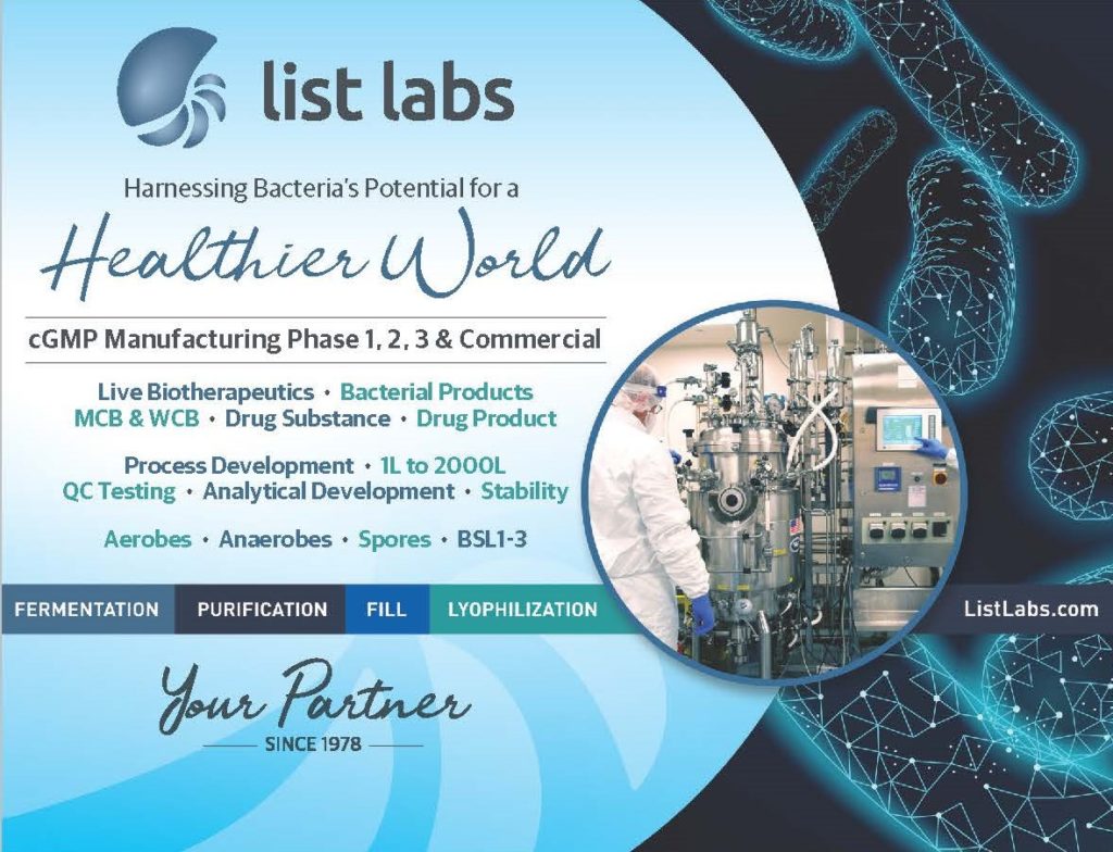
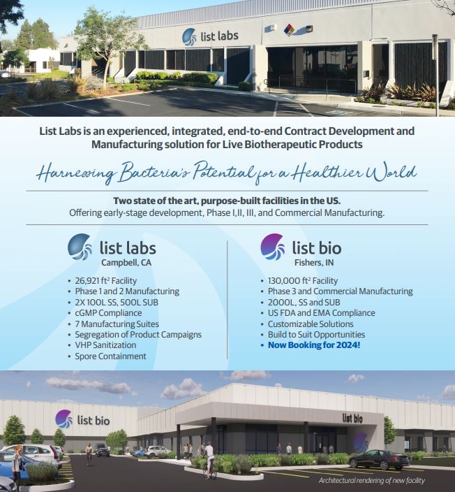

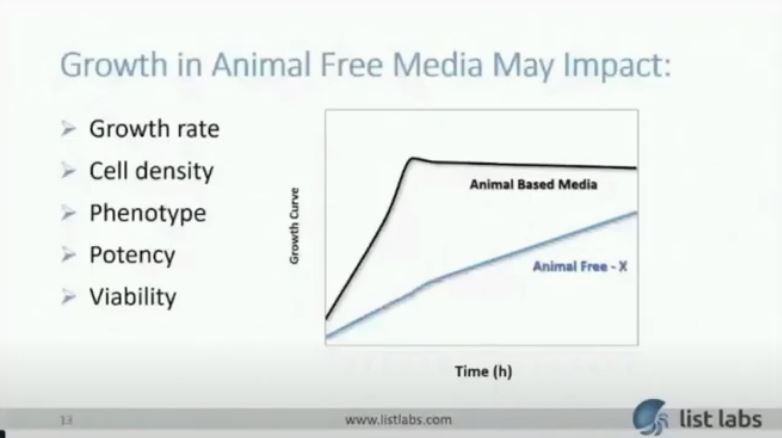


 Microbiome research is uncovering the enormous potential for developing drugs, such a live biotherapeutic products, from the microbiome. This burgeoning field is the future of medicine.


 List Labs has extensive experience with microbiome and more specifically

 List Labs
 By: Rachel Berlin, Marketing Manager
 By: Rachel Berlin, Marketing Manager


 Find a distributor near you